Surgical Approach to the Pseudoaneurysms Of Lower Extremity Arteries Developed After Gunshot Injuries
U Yetkin, S Bayrak, � Tetik, B Lafç?, C Özbek, M Ye?il, A Gürbüz
Keywords
gunshot injury, lower extremities' arteries, post-traumatic pseudoaneurysm, revascularization
Citation
U Yetkin, S Bayrak, � Tetik, B Lafç?, C Özbek, M Ye?il, A Gürbüz. Surgical Approach to the Pseudoaneurysms Of Lower Extremity Arteries Developed After Gunshot Injuries. The Internet Journal of Thoracic and Cardiovascular Surgery. 2006 Volume 10 Number 2.
Abstract
Background and Aims: Trauma is the third leading cause of death in the general population and the leading one among people younger than 45 years. Vascular injuries comprise 3% of the traumas in the social life. The most important late-period complication due to gunshot injuries of lower extremity arteries is pseudoaneurysm. This study was undertaken to describe the management of pseudoaneurysms (PSAs) of lower extremity arteries after gunshot injuries.
Material and Methods: Between January 2001 and December 2006, we performed revascularization for PSA in the femoral, popliteal and tibioperoneal lower extremity arteries of eight patients who had gunshot injuries. There were 8 male patients with mean age of 30.1 years (range: 24 to 37). All patients underwent color-flow arterial Doppler ultrasonography and selective lower extremity digital subtraction angiography.
Results: The median interval between injury and presentation was 18 weeks (range 6 weeks to 9 months). In all patients, we performed aneurysmal resection and all patients were treated with optimal revascularization procedure. It was determined that one patient operated by us due to popliteal artery developed graft occlusion at the postoperative 6th hour. We re-explored this patient and performed a successful embolectomy. All patients were able to completely straighten the leg at the time of discharge. The mean follow-up duration was 1.9 years (range, 3 months to 6 years), and the mean time to discharge was 5.3 days (range 5-7 days). No deaths or graft related complications occurred in these patients and the early and late patency and limb salvage rates were 100%.
Conclusion: Early diagnosis of the PSAs in lower extremity arteries constitutes an important factor in successful surgical reconstruction. Open surgical repair must be used as the standard approach to symptomatic and rapidly enlarging PSAs in order to avoid rupture, thrombosis and embolization, which threaten the function and vitality of the extremity, and less invasive methods must be reserved for rare and complicated cases.
Introduction
Trauma is the third cause of death in the general population and the leading one among the people younger than 45 years. Vascular injuries comprise 3% of the traumas occurring in the daily life and even in 21st century, morbidity and mortality rates are significantly high. Acute or chronic traumas may cause severe peripheral vessel lesions (1). Gunshot wounds affecting the main vessels of the extremities mostly threaten limb salvage (2). Lower extremity pseudoaneurysms develop after penetrating traumas lead to direct vessel injuries. 64% of all arterial injuries and 78% of all vascular injuries are gunshot wounds (3). Extremity vessels are affected in 75% of them. Especially in wounds with high speed gunshot; endothelium, adventitia and elastic laminas are injured and late thrombus and pseudoaneurysm develop (4). A penetrating trauma, directed to vessel wall, can cause an acute traumatic pulsatile hematoma by rupturing all the layers of arterial wall (5). Hematoma is limited with adjacent tissues or organs and enlarges until it is surrounded by a chronic fibrous wall or rupture. Chronic traumatic aneurysm develops by the partial or complete absorption of periarterial hematoma in the fibrous sac, which is surrounded with adjacent tissues. Generally the term “traumatic aneurysm” is used to refer to pseudoaneurysm (5,6). Regardless of whether they are acute or chronic, pseudoaneurysms (PSAs) must be treated as soon as possible after the diagnosis is made with direct surgical procedure. Rupture risk, thrombosis and compression to adjacent structures can be avoided by early operation (5). In this study, we present 8 patients with pseudoaneurysms localized at three different levels (femoral, popliteal and tibioperoneal) of lower extremity arteries, which developed after gunshot wounds and our surgical repair approach with review of literature.
Material And Methods
Between January 2001 and December 2006, we performed revascularization for PSA in the femoral, popliteal and tibioperoneal lower extremity arteries of eight patients due to gunshot injuries in our Cardiovascular Surgery Department. There were 8 male patients with mean age of 30.1 years (range 24 to 37 years). First aid was rendered to these patients at other medical institutions. There was no pulse deficit or ischemia sign and their clinical pictures and hemodynamics were stable at first admission.
We examined the lower extremities for ischemia, the patients were questioned verbally about their medical history and physical examination was performed. All these procedures contributed to the diagnosis. We classified our patients into three groups: Group I consisted of two patients with femoral artery PSA, Group ll consisted of four patients with popliteal PSA and Group III included two patients with tibioperoneal PSA (Table 1).
Figure 1

All patients presented with a pulsating mass at the affected arterial level, which had developed in the late post-traumatic period (Figure 1).
Figure 2

The patients were admitted to our clinic for progressive tumor, function loss and pain. Their affected legs were slightly cold and peripheral pulses could hardly be palpated on the affected legs of the patients compared to the unaffected legs. There was pulsation on the mass and murmur correlated with systolic thrill was heard in all patients. Ankle/arm index was 0.7-0.8 at affected leg and ≥1 at uneffected leg. Except a hematocrit value of 26% determined in one of our patients in Group I (Patient no.2); routine biochemical tests, the results of the bleeding and coagulation timing tests were normal in all patients. Lower extremity arterial and venous colored Doppler ultrasonographic (CDUSG) tests revealed a giant hematoma measuring 6-20 cm diameter and hemorrhagic liquid areas with various concentrations at the localizations of PSAs (Figure 2).
Figure 3

Saccular formations measuring between 4 and 15 cm diameter were determined in all PSA patients and the “to and fro” flow pattern was thought to indicate pseudoaneurysm (Figure 3).
Figure 4

No patient had an arteriovenous fistula. One patient in Group lll (Patient no.8) presentesd with an arterial pseudoaneurysm under the tibioperoneal truncus, complicated with pseudoarthrosis sequela secondary to tibial osseous fracture and superficial and deep venous chronic thrombophlebitis (Figures 4 and 5).
Figure 5

Figure 6

The venous structures of the remaining 7 patients were normal. An important anamnestic finding was already present in patient no.8 before he was admitted to our clinic. Invasive radiology unit planned coil embolization for distal left tibioperoneal truncus occlusion and left crural pseudoaneurysm but as the procedure failed and the symptoms increased, the patient was referred to our clinic. We performed selective lower extremity arteriography with digital substraction angiography (DSA) in all patients (Figures 6, 7 and 8).
Figure 7

Figure 8
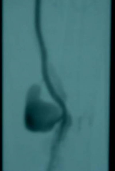
Figure 9

Each patient underwent surgery after the diagnosis of late post-traumatic lower extremity arterial pseudoaneurysm was made.
Results
The diagnostic delay since injury ranged from 6 weeks to 9 months, with a median delay of 18 weeks. The patients in Group I were operated under endotracheal general anesthesia, in supine position. Bupivacaine was used for spinal anesthesia in patients in Group II and III. The surgical procedures that we performed in 8 patients are presented in Table 2.
Figure 10

Generally, we used the following principles in the surgical procedures that we performed in our patients: aneurysm was determined with skin incision parallel to the affected artery course and exploration was carried out to determine whether adjacent vein and nerve structures were damaged besides the injured artery. Saphenous vein grafts were prepared from the vena saphena magna of the contralateral legs in four patients. Saphenous vein graft was used in four patients (Patients no. 1, 3, 4, 6 and 7). After administering 1cc heparin (5.000 İU) intravenously, bleeding was controlled with vascular clamps and the capsule of the pseudoaneurysm was opened with direct incision. The organized thrombus in the aneurysm was removed (Figure 9).
Figure 11

In order to avoid the risk of major hemorrhage or nerve injury, we did not resect the aneurysmal pouch completely. We performed limited the resection by preserving the adjacent tissues. Embolectomy performed with distal 3 Fr Fogarty catheter revealed materials only in one patient (patient no.8) The aneurysm of this patient was successfully removed with distal-proximal ligation. The diameter of the saphenous vein graft was not enough for interposition in patients no:2 and 4, so patency was constructed with 6mm ringed expanded polytetrafluoroethylene (e-PTFE:Gore-tex) tube graft interposition (Figure 10).
Figure 12

The capsule was dissected and evaluated histopathologically and microbiologically. A closed drainage system was placed in the sac of giant aneurysm and and the aneurysm sac was closed with endoluminal circular sutures. Except one patient (patient no.7), all distal pulses were similar to the opposite ones during postoperative period. Distal pulse disappeared at the postoperative sixth hour in this patient and he was re-explored and a successfull embolectomy was performed with a 3 Fr Fogarty catheter. No complications developed during the late postoperative period. His popliteal artery was primarily reconstructed and the examination performed via lower extremity DSA at the postoperative 2nd month showed that the primary reconstruction was successful.
Low molecular weight dextran (500 ml / day), pentoxyphylline (300 mg/day) and anticoagulant (25.000 U/day unfraxioned heparin with ACT/12h control) were administered postoperatively for 3 days. The microbiologic examination showed that aneurysmal capsule was negative for microorganisms. Histopathologic evaluation of the pseudoaneurysmal capsules showed that they were all hematomas with extravasation in the vessel wall lumen, inflammatory infiltration and hyalinization. Histologically, the well encapsulated aneurysmal sac lined organized hematoma and the arterial wall was replaced by fibrous tissue (Figure 11).
Figure 13

The elastic stain also did not reveal any elastic fiber (Figure 12).
Figure 14

In all patients a pulsation was positive upon digital examination at the distal anterior tibial and posterior tibial arteries during the early postoperative period. The late postoperative follow-up examinations were performed at our clinic and Doppler ultrasonography was performed after 3 months.
The early (first month) and late graft patency rate was 100% and we found no other sequelae during the late period. The mean follow-up was 1.9 years (range, 3 months to 6 years), and the mean time to discharge was 5.3 days (range 5-7 days).
Discussion
Vessel trauma can cause systemic, regional and local pathophysiological problems. Local and regional reactions are determined by the vascular trauma type (1). The mechanism of injury is gunshot in 40% of the cases (7). The most destructive effects are seen on vessels due to the kinetic energy of the bullet (1). The most frequently injured vessels are femoral arteries (37.3%), followed by the popliteal (27.8%), axillary and brachial (23.5%), and crural arteries (6.5%) (7). Injuries without acute problems may lead to pseudoaneurysm and A-V fistula in time (1). PSAs of the lower extremity arteries are very rare and occur as a late complication after arterial injury (8). In Vietnam War, pseudoaneurysm developed in 7% of all vascular injuries (6). It should be kept in mind that patients with trauma history and pulsatile masses may develop pseudoaneurysm (5,9). Complication factors are amount of energy causing injury, wound infection, nevre injury, compartment syndrome, malunion of fractures (1,10). Osseous fractures of patient no.8 constitute an example for this.
Generally, extremity vessel injuries are seen in young men, aged between 20 and 40 (1). As seen in Table 2 all our patients were men in this age period.
Even with severe arterial injury, ischemia signs cannot be seen distal to injury (11,12). It has been reported that distal pulses can be palpated especially in patients without hypotension and with developed collateral vessel system, even if arterial tear is present (11). In a study of Peck, distal pulses could be palpated in 10% of the patients with severe popliteal arterial injury (13). Palpated pulse can be felt with intimal flap, smooth fresh thrombus or collaterals (1,12). In some of the vessel injuries, significant injury findings are not present and necessary prevention measures are not taken. Thus, serious complications and even death may occur. Therefore, these patients must be closely examined if injury is proximal to large vessels and if there is any suspicion; arteriography must be performed (1). Angiographic intervention is recommended for gunshot and especially for buckshot injuries because more than one vessel may be involved (14). We performed angiography in all patients.
Clinical signs of pseudoaneurysms include pulsatile mass, murmur over its localized region, palpable thrill, pain and extremity edema (15). Our patients had all these symptoms. Pseudoaneurysms following trauma cause pain by compressing the adjacent structures, can contain thrombus and can cause regional ischemia due to distal embolus(1). Typically pseudoaneurysms do not prevent blood flow along the natural vessels. So, distal perfusion is protected. The most important risk is blood loss from outside of the sacculus, which occurs due to rupture (16). Additionally, chronic symptoms secondary to compression resembling chronic trombophlebitis, may be present in patients like Patient no.8 (17).
Color – flow Doppler ultrasonography (CFDU) is a noninvasive method that can provide sufficient diagnostic information to plan the surgical procedure (18). CFDU has an excellent sensitivity and specificity spectrum and shows the blood filling the cavity as well as the jet flow passing through the pseudoaneurysmal sac with arterial defect (18,19). Its advantages are low cost, utility in various treatment choices and low time consumption. In addition to its use in making the diagnosis, it provides detailed information about the dimensions, morphology and neck anatomy of the pseudoanurysms, the blood flow through the pseudoaneurysms and the relation of pseudoaneurysms with the adjacent vessels (15). Lower extremity arterial Doppler ultrasonography and magnetic resonance angiography can be used as diagnostic tools, but the gold standard is selective lower extremity arteriography (20). Doppler ultrasonographic evaluation is sufficient for late postoperative follow-up evaluation (18,19).
Therapeutic interventions include open surgical repair, ultrasound guided compression, thrombin injection under ultrasound, coil embolization and endovascular repair with stent-graft (1, 12, 15).
Endovascular repair methods are not used routinely for femoral pseudoaneurysm treatment. Generally, they are preferred when conventional methods are too risky or technically impossible to be used (8). Although it is technically possible to place stent for femoral or popliteal pseudoaneurysms, its patency rate is between 43% and 87%, which is not satisfactory (15, 21, 22). Tool migration or fracture and endoleak are complications specific to to stent-graft repair and can be improved by stent excision with open repair (22).
Coil embolization, which failed in patient no.8, does not have a wide field of utilization in peripheral pseudoaneurysms. It is mainly used for cerebral vascular aneurysms, A-V malformations, traumatic or other etiologic visceral and pelvic hemorrhages, A-V fistulas or in neoplasm treatment for reduce blood flow, obliteration of unligated a.mammaria interna lateral branch in mammarian steal syndrome after coronary by pass surgery and obstruction of saphenous lateral branches during in situ saphenous by pass (23). Embolization of pseudoaneurysms can be achieved only by embolization of the sac: the pedicle of the sac must be small, and the aneurysm must not disturb the distal circulation to a great extent. Another method -embolization of the artery from the distal and proximal arterial segments- can be used if collateral circulation is sufficient (24,25).
Open surgical repair should always be the first choice of treatment (15,26). Traditional surgery is still considered to yield the best results. For surgical reconstruction of late complications of arterial injuries such as fistula and pseudoaneurysm, end to end anastomosis is the first choice of treatment and when it is not possible to perform end to end anastomosis, saphenous vein graft interpositioning in recommended (3,27). Today, many clinics prefer autogenous grafts which are more resistant to infection and have an elongated patency for extremity vessel injuries (20,28). As for our Group I patients, resection + graft interposition was the simplest and successful method in open repair. The most convenient graft material to be chosen in patients such as patient no.2 is PTFE. Autogenous vein grafts have advantages only if there is infection (8,13). 5-year patency rate for open repair of femoral arterial aneurysms is 85% (29).
Autogenous grafts must particularly be used for popliteal and infrapopliteal injuries because the risk of thrombosis is high in synthetic grafts with a diameter less than 6 mm (20,28).
In many patients with penetrating injuries, which occur as a result of the violence in the social life, tibial artery injuries are treated with ligation and this treatment yields successful results. As for tibial artery injury, ischemic late complications are not seen after ligation if the distal leg is not ischemic, (27). Distal and proximal ligation of the pseudoaneurysm and resection can be applied in patients with lesions in distal arteries, which are not susceptible to cause severe ischemia after the intervention and those patients who are previously confirmed to have sufficient collateral circulation after the ligation (23). If aneurysm is solitary and distal to tibioperoneal truncus and has a limited diameter, it can be ligated but if the pseudoaneurysm includes tibioperoneal truncus or if it develops in multiple and more distal arteries, reconstruction techniques are necessary in order to assure the viability of the extremity (23,30).
We also preferred a medial surgical approach to the leg, because it is easy to perform and when the patient is supine position it offers many choices. Besides tibial vessels, vena saphena manga can also be exposed easily with this method. It is very important to reach posterior and peroneal vessels for distal artery by pass (31). Additionally, if the situation of the popliteal artery located at the upper patella level is worse than expected, this approach provides enough inflow from superficial or common femoral artery.
As a conclusion; today, vessel injuries and late-period complications keep increasing in parallel with the increase in violence and crime in the social life. Physical examination is not enough for perforating injuries directed to extremity arteries. Thus, noninvasive and invasive investigations must be used to exclude possible complications if necessary. As a late-complication, the pseudoaneurysm must be repaired with traditional open surgery to prevent the extremity function and vitality. We recommend surgical reconstruction techniques for the repair of lower extremity pseudoaneurysms. In addition to the surgical procedure, interventional radiological techniques such as endovascular intervention and embolization can be used for the treatment of pseudoaneurysms. They have advantages such as of low blood loss, reduced anesthesia requirement in patients with poor general health and centrally located pseudoaneurysms. We recommend that open surgical repair should be performed as the standard approach and if necessary selective revascularization should be used together with the routine surgical repair of aneurysms.
Correspondence to
Doç. Dr. Ufuk YETKİN 1379 Sok. No: 9,Burç Apt. D: 13 35220, Alsancak – İZMİR / TURKEY Tel: +90 505 3124906 Fax: +90 232 2434848 e-mail: ufuk_yetkin@yahoo.fr