Gunshot Wounds Sustained Injuries To The Face: A University Experience
C Perry, B Phillips
Keywords
medicine, plastic surgery
Citation
C Perry, B Phillips. Gunshot Wounds Sustained Injuries To The Face: A University Experience. The Internet Journal of Surgery. 2000 Volume 2 Number 2.
Abstract
Introduction
Missile injuries to the face usually present in dramatic fashion and demand clinical savvy for patient survival. A definitive airway is often required in this population and appropriate wound management is imperative for an acceptable outcome. Offered is an illustrative case of a self-inflicted gunshot wound to the lower face followed by a retrospective review of our University experience. Given the pattern of injury typically associated with this mechanism, initial airway management and definitive reconstruction options are discussed.
Case Report
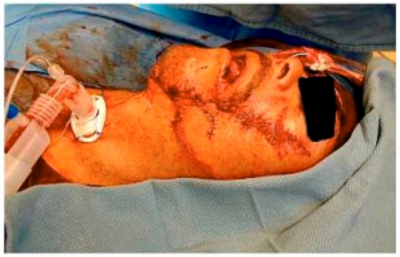
A forty-four year old male with massive facial trauma from a self-inflicted wound presented to the emergency department.[Figures 1 & 2]
Figure 1

Figure 2

A nasal endotracheal tube was inserted in the field for airway protection and its position was confirmed by auscultation. He was then air-lifted from his hunting camp to our Level I trauma center. On arrival, a pulse of 80 beats per minute, a blood pressure of 170mmHg over 60mmHg, and an oxygen saturation of 75% were obtained. An initial arterial blood gas revealed a pH 7.34, a carbon dioxide level of 47mmHg, and a partial pressure of oxygen of 35mmHg. A lateral head and neck radiograph revealed a nasal esophageal tube and multiple foreign body fragments.[Figure 3]
Figure 3

The nasally-placed was removed and the patient was successfully orally intubated using lidocaine and sedation without paralysis. Tube position was confirmed with directed laryngoscopy, capnography, and a chest radiograph. Subsequent physical examination revealed a left facial nerve palsy of the mandibular, buccal, and zygomatic branches. The patient remained hemodynamically stable throughout his emergency department course. Computeized tomography of the head and facial bones revealed loss of bony integrity including fractures of the mandible, zygomatic arch, orbital floor, and maxilla with multiple metallic fragments throughout the soft tissues. There was not an appreciable intracranial injury. An angiogram of the neck confirmed internal carotid vessel integrity while revealing a lacerated facial artery. He was taken to the operating theatre for tracheostomy, debridement, and closure of the facial laceration. His mid-face fractures were subsequently plated and a bar was placed to bridge the bone loss of the mandibular body.[Figure 4]
Figure 4
The patient has required serial debridements but continues to do well while awaiting definitive bone grafting and coverage.
University Experience: Methods
A retrospective review of the trauma registry from January 1, 2000 to December 31, 2000 at Tucson medical center was performed in order to identify patients sustaining missile injuries to the face. 1,156 trauma patients were seen during this time period and 692 were admitted. Of these, 119 patients were immediately taken to the operating room for a variety of surgical procedures. There were a total of fifty-six gunshot wounds identified – fourteen of which involved the face.
Results
Fourteen (25%) of the fifty-six patients identified with gunshot wounds sustained injuries to the face.[Table 1]
Figure 5

Of this sub-group, there were ten males (71%) and four females (29%). The average age of all patients was 34 years. Six (43%) of the injuries were self-inflicted, aimed directly at the anterior face. Of the fourteen patients, three (21%) died secondary to their injury; all deaths were from self-inflicted wounds. Six patients (43%) required an urgent surgical airway. There were eight (57%) intracranial injuries, three (21%) facial nerve injuries, one (7.1%) carotid injury, one (7.1%) parotid gland injury, and one (7.1%) patient that sustained multiple facial fractures secondary to the missile tract. Of important note, none of our patients sustained a cervical spine injury- despite four patients having missile tracts though the neck-proper. Eight patients (57%) required serial operative debridements followed by plastics reconstruction. Four (29%) tracheostomies were performed as well as two cricothyroidotomies (14.2%). One (7.1%) patient required open reduction and internal fixation of multiple facial fractures and a second patient underwent a facial nerve graft.
Discussion
There are four main steps in the management of patients with gunshot wounds to the face: securing an airway, controlling hemorrhage, identifying other inuires, and definitive repair of the traumatic facial deformities.[Table 2]
Figure 6

Airway management can be difficult as exemplified by our case report (an esophageal intubation). In the medical literature, an urgent airway is required in up to 35% of patients presenting with these injuries.1 In up to 83% of patients, oral endotracheal intubation is initially succesful2, though our university experience was far different. A surgical airway is required in the remaining 17% of patients with facial trauma secondary to gunshot wounds.3 This is usually due to tracheal occlusion from edema, loss of bony integrity, or active hemorrhage. Airway control in patients with severe mid0facer trauma has been well-described in the medical literature.4 [Table 3]
Figure 7

Fiberoptic intubation without paralysis is becoming a more-practical option for some of these patients. Blind nasal intubation is contraindicated in patients with basilar skull fracrtures, CSF leaks or mid-face instability; there are multiple reports of nasally intubating the cranium in such patients.5 The enxt vailable option when faced with this mechanism of injury, is an oral tracheal airway, however, it is imperative that the trauma team be prepared for a cricothyroidotomy if the airway can not be quickly controlled. Needle ventilation of the trachea followed by tracheostomy is another surgical option. Direct layngoscopy, capnograohy, and a chest radiograph are all useful to confirm placement as well as to evaluate possible foreign body aspiration. After the patient has a secure airway and bilateral breath sounds have been confirmed, hemostasis then takes priority. While both angiography and directed surgical control are options for hemostasis, angiographhy may be the preferred method and is helpful in the evaluation of anterior neck trauma (e.g. zones I and III).6 After the hemorrhage is controlled either by direct pressure, packing, embolization, or direct ligation, the patient can be resuscitated and evaluated systemically to “rule-out” other injuries.
The secondary physical examination of the face must consider the entry point of the missile, velocity, trajectory, and exit site. These factors, if easily discerned, may have some prognostic significance.7 The laceration size, depth, and location to the neurovascular supple with surrounding tissue injury must also be taken into account. Facial fractures should be identified with a thorough and complete examination followed by plain radiographs. Computed tomography of the brain, face, and neck will assist in the evaluation of associated injuries as well as to quantify the extent of facial fractures - both axial and coronal images should be obtained to assist in reconstructive management. However, the cervical spine must be completely “cleared” prior to obtaining coronal images. In the literature, gunshot wounds to head and neck will have intracranial parenchymal involvement in approximately 17% of patients and a cervical spine injury in 8%8
The most common bony injury, in order of occurrence, associated with gunshot wounds to the face include the mandible, maxilla, and zygomatic arch.9 Approximately one-third of patients will require operative debridement followed by fixation.10 In our experience, 57% of patients required serial debridements. Only after the full extent of the injury has been appreciated and allowed to “demarcate”, can reconstruction be considered.
Attempts at immediate definitive reconstruction often fail secondary to loss of mucosal integrity and septic necrosis of the surrounding bone. Traditionally, these wounds were managed with conservative debridement, serial dressing changes, external fixation, and delayed reconstruction. This style of management required social isolation of the patient for months, scar contracture, and bacterial colonization of the wound. Optimal cosmetic forma and function can be obtained with a more-timely surgical approach. This requires the surgeon to account for an overall increased risk of infection due to tissue destruction associated with the missile tract, creation of dead-space, retained foreign bodies, and entrapped oral flora. A threes-staged approach has recently been championed by Manson.11 This first stage involves anatomic definition of the tissue and bone loss. After this is quantified by physical examination and computerized tomography, conservative irrigation and debridement can follow systemic stabilization of the patient: serial explorations are the rule rather than the exception. The second general step in mangement is wound stabilization through restoration of bony relationships. Dental occlusion is properly established by dividing the face into two halves. The lower half consists of the mandible and maxilla, while the upper half is comprised of the maxilla, orbits, and zygomatic process. Once the occlusal relationship between the maxilla and mandible are optimally established, the upper facial fractures are then repaired. Establishment of an appropriate skeletal buttress allows maximal preservation of function and cosmesis. Early reconstruction provides optimal skeletal stabilization and skin coverage preservation. The key relationships of the upper half of the face include the periorbital rims, zygomatic arches, and the zygomaticomaxillary struts. Bony loss is often bridged initially with metallic struts atytempting to prevent soft tissue contracture or collapse with devascularized bone grafts.
Computed tomography is often helpful in this stage, helping to maintain a proper three-dimensional relationship. Once projection is restored, height and overall facial width have been addressed. The wound is then re-explored at various intervals (24 – 36 hours) to debride the devitalized tissue, remove any subsequent hematoma, and insure mucosal integrity. Ideally, the integrity of the mucosal lining can be obtained within 5-7 days of the initial injury. Repairing the face by functional and aesthetic units allows for optimal healing. Bony loss in the upper face is treated by skeletal splints and devascularized bone grafts. These devascularized bone grafts require obliteration of dead space, intact epithelial and mucosal coverage for viability. The lower facial structures will often require the replacement of entire facial units in order to restore bony and cutaneous functional elements. Microvascualr tissue transfers are often utilized in this situation. Patient follow-up in this population has traditionally been poor but in those that are compliant, secondary interventions may be performed for debulking, color modification, and scar revision.
Conclusion
Although our patient’s oputcome was unaffected by the initial failed airway, this case illustrates the importance of ensuring a definintive airway in this subset of trauma patients. Capnography confirmation or direct laryngoscopy in the field might have detected the misplaced airway earlier in his course. In our review of the medical literature, up to forty percent of these patients will require an urgent airway and up to twenty percent will require a tracheostomy. It is interesting to note that of the deaths occurring in our experience, all were associated with self-inflicted wounds. Once the patient has been resuscitated, other injuries must be identified and addressed in the standard ATLS manner. It is important to then develop a reconstructive surgical plan after debriding devitalized tissue. Restoration of facial integument in an expeditious manner will help avoid disfiguring morbidity associated with contracted facial wounds.
Correspondence to
Charles W. Perry, M.D.
The University of Arizona
Dept. of Surgery
1501 North Campbell Ave.
P.O. Box 245058
Tucson, AZ 85724-5058
Fax: (520) 626-2247
Phone: (520) 626-7747