Anasarca and Orophayngeal Myopathy: Unusual manifestations of Polymyositis
T Trung Phan, M Asante-Abedi, A Faizal, S Bain
Keywords
muscle atrophy, polymyositis, subcutaneous oedema
Citation
T Trung Phan, M Asante-Abedi, A Faizal, S Bain. Anasarca and Orophayngeal Myopathy: Unusual manifestations of Polymyositis. The Internet Journal of Rheumatology. 2005 Volume 2 Number 2.
Abstract
We present a case of polymyositis affecting a 56 years old man in whom the predominant presenting features were myalgia and anasarca (generalized oedema) of the upper limbs and thorax. The patient went on to have involvement of the lower limb, respiratory and pharyngeal musculature, the latter leading to an aspiration pneumonia and prolonged nasogastric feeding. Despite this stormy course, he has made a good recovery and his long-term prognosis is good.
Introduction
Polymyositis was first recognised in 1887 by Wagener and Unverricht in patients presenting with muscle weakness and skin lesions. In 1958, Walton and Adams described the additional features of pain, arthralgia, fatigue and fever. Nowadays, the commonly used diagnostic criteria are: symmetrical weakness of limb girdle muscles worsening over weeks-to-months; raised serum creatine kinase; electromyographic changes; and muscle biopsy abnormalities. (1)
The prevalence of polymyositis is estimated to be 1 per 100,000. (2) Presentation is bimodal and is more common in women (2:1 ratio). The peak age is 10-15 years for children and 45-60 years for adults. Mean age varies according to the subset. We describe here a male patient who presented with polymyositis associated with profound generalized subcutaneous oedema complicated by muscle atrophy and oropharyngeal muscle paresis.
Case Report
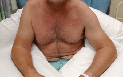
A previously fit 56 year old man presented with severe pain and swelling affecting the upper limbs and anterior chest wall. He gave a 7-week history of malaise, fatigue and progressive muscle weakness associated with arthralgia. He smoked 20 cigarettes per day and had previously taken excessive alcohol. He had no other history and was not taking regular medication. At presentation, there was marked oedema of the upper limbs and anterior chest wall; palpation of these areas induced severe pain (see Figure 1). There was also weakness affecting the proximal leg muscles, such that he had difficulty standing unaided, but no associated oedema or tenderness. There was no involvement of the swallowing, neck and extra ocular muscles.
Figure 1
Full blood count, urea, electrolytes, erythrocyte sedimentation rate and thyroid function tests were all within normal limits.
In view of the marked oedema, CT thorax was performed to rule out superior vena caval obstruction. This was normal, and also showed no evidence of malignancy. CT scan of the abdomen was normal and tumour markers were not elevated. On the third day of admission, muscle biopsy was performed and showed localised perivascular lymphocytic infiltrates and mild fat replacement suggesting fibre loss. This was thought to be consistent with a necrotising myopathy likely to be inflammatory in nature.
Treatment was commenced with prednisolone (60mg OD) and bone protection using alendronate (75 mg/week). After one week, creatinine kinase fell to 2430 U/L, however, the clinical picture worsened; oedema and muscle tenderness became generalised with involvement of the lower limbs. Subsequently he was noted to be breathless with reduced respiratory excursion and he developed dysphagia with reduced gag reflex. On day 10, he developed an aspiration pneumonia requiring ventilation and inotropic support. At this time, an echocardiogram showed good left ventricular function. He made a full recovery from the pneumonia but continued to have poor chest muscle strength and an absent gag reflex. In view of the high risk of further aspiration, he remained on nasogastric feeding until the gag reflex was clearly present, a period of ten weeks.
After 3 weeks of steroid treatment, the oedema began to reduce, but he was left with generalised muscle wasting (see Figure 2). Once the creatinine kinase had normalised, prednisolone was tapered down by 5mg every week and he was started on 50mg of Azathioprine daily, gradually increased to 150mg.
Figure 2

Discussion
Gross anasarca is an unusual presentation of polymyositis. Initially, this was most noticeable in the upper limbs and anterior chest wall but over 48 hours spread to involve the lower limbs. This feature has only been reported in a small number of case reports. (3,4,5) The mechanism of the generalized oedema is likely due to an increase in capillary permeability as a result of the intense inflammatory processes in the underlying muscle. The patient also experienced severe myalgia and muscle tenderness which is typical in 30% of patients with polymyositis. Whilst muscle weakness is a common feature of polymyositis, muscle atrophy is relatively rare so early in the course of disease. This feature became more obvious as the oedema settled. Seldomly, muscle weakness can also involve the oropharyngeal muscles and the striated muscle of the upper third of the oesphagus. (6) This can cause dysphagia leading to increased risk of aspiration pneumonia as was seen in this case.
The clinical diagnosis of polymyositis can be confirmed by three tests: serum muscle enzymes, electromyography and muscle biopsy. Muscle enzymes such as creatine kinase (CK), lactate dehydrogenase (LDH), aldolase, aspartate aminotransferase (AST) are typically raised during the course of the illness but can be normal. (7) A correlation between the rise in muscle enzymes and severity of weakness has been reported (8), however, other studies have found that treatment response and disease progression bare little relationship to the CK levels. (9) In this particular case, the elevation of CK correlated best with the extent of the oedema, on the other hand, muscle weakness and myalgia persisted long after the CK had fallen back to normal levels.
Polymyositis is an inflammatory myopathy in which the pathogenesis is presumed to be autoimmune. It is believed that CD8–positive cells invade MHC–I-antigen expressing muscle fibres leading to fibre necrosis.(10) The auto-antigen has yet to be identified. Autoantibodies, such as antinuclear antibodies present in this case, are found in 80% of patients with polymyositis and dermatomyositis (DM) (11), however they are usually non- specific.
Muscle biopsy is considered to be the definitive test for polymyositis and it also allows for exclusion of other myopathies. Ideally the biopsy should be taken from the contralateral electromyogram (EMG) proven group of muscle or from the involved muscle group identified by magnetic resonance imaging. Histological features include muscle fibre degeneration and necrosis, and an inflammatory cell infiltrate. Some of these features were observed in the muscle biopsy of this case. A more distinct histological feature is cellular infiltrate within the fascicle.
One of our concerns in this case was the possibility of an underlying malignancy because patients with polymyositis has a 2-fold increase in the frequency of malignancy. (12) The most common cancers involve: ovaries, bowel, breast, lung and non-hodgkins lymphomas Fortunately the CT scans and tumours markers were all negative at the time and no evidence of malignancy at 12 months follow-up.
The mainstay of treatment of polymyositis is corticosteroids. As yet there are no randomized controlled trials to show that this therapy has an impact on survival but studies have shown benefits in preservation of muscle strength. (13) In patients who respond, CK levels should normalize within 4-6 weeks, however, muscle strength can take 2-3 months to improve. Azathioprine and methotrexate can be used in conjunction with prednisolone at the beginning or later in patients with incomplete response to corticosteroids. Intravenous immunoglobulin has also been shown to have some benefits. (14)
Overall, the prognosis of polymyositis is good with a 5-year survival of 95% and10-year survival 84%. Poor prognostic factors include increasing age, duration of illness prior to diagnosis, severity of weakness, dysphagia, and recurrent aspiration pneumonia. (15)
Learning Points
-
Myalgia and gross generalised oedema can be the presenting signs in polymyosities
-
Polymyositis and its complications such as weakness of oropharyngeal muscles leading to dysphagia and life threatening risk of aspiration pneumonia should be recognized early and managed effectively.
Correspondence to
Thanh Trung Phan MRCP Clinical Research Fellow University of Birmingham Department of Cardiovascular Medicine Edgbaston Birmingham B15 2TT, UK Tel: 0121 424 1771 Fax: 0121 424 1074 E-mail: ttpquang@hotmail.com