Pulmonary nocardiosis presenting as bilateral pneumonia in an immunocompetent patient – An unusual host response
S Kumar, R Pajanivel, N Joseph, S Umadevi, M Hanifah, R Singh
Keywords
granulomatous response, lung, nocardia, pneumonia
Citation
S Kumar, R Pajanivel, N Joseph, S Umadevi, M Hanifah, R Singh. Pulmonary nocardiosis presenting as bilateral pneumonia in an immunocompetent patient – An unusual host response. The Internet Journal of Microbiology. 2009 Volume 9 Number 1.
Abstract
Pulmonary nocardiosis (PN) is an infrequent and severe infection due to
Introduction
Pulmonary nocardiosis is an infrequent but severe infection that commonly presents as a subacute or chronic suppurative disease, mimicking a lung carcinoma, abscess or pulmonary tuberculosis (Gillespie, 2006).
Case report
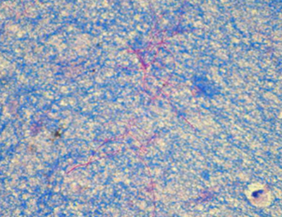
A 24-year-old female was brought to the casualty with complaints of breathlessness, abdominal pain and vomiting for six days. She had productive cough with purulent sputum mixed with blood for 12 days. She was apparently normal two weeks back. There was no evidence of immunocompromised status. On admission, she was conscious, oriented, febrile and tachypneic. Her pulse rate was 126/min, temperature 39.1°C (102.4°F), respiratory rate 32/min, and blood pressure 110/80 mmHg. Chest auscultation revealed bilateral crepitations. Her hemoglobin was 8.6 g/dL and the leukocyte count 16,000 cells/ cu mm with 2% bands, 74% neutrophils and 24% lymphocytes. Microbiological examination of the sputum failed to identify a pathogen. A chest radiograph revealed bilateral irregular nodules (cavitating) with indistinct areas of haziness, prominent broncho-vascular markings and mild effusion (Figure 1). Abdominal examination was normal. Based on clinical, laboratory and radiological investigations a provisional diagnosis of community-acquired pneumonia was made and the patient was put on meropenem. Despite broad-spectrum antimicrobial therapy, her condition deteriorated. A pleural tap was done. Cytological smears of the pleural effusion demonstrated numerous neutrophils and reactive mesothelial cells. Microbiological examination of the drained fluid revealed Gram positive filamentous and branching bacilli, which was weakly acid fast by modified Ziehl Neelsen staining, suggestive of
Figure 1
Figure 2
Discussion
Nocardia is an uncommon pathogen in immunocompetent patients; however, it has been increasingly recognized as a significant opportunistic pathogen in immunocompromised individuals. Pulmonary disease is the predominant clinical presentation (more than 40% of reported cases), with almost 90% of these caused by members of
Conclusion
Pulmonary infection by this pathogen may thus be difficult to diagnose based on clinical and radiological features as these are not specific. Nocardiosis should always be considered in the differential diagnosis of indolent pulmonary disease even in immunocompetent patients. Our case illustrates the need for a high index of suspicion of pulmonary nocardiosis. Although pulmonary nocardiosis is usually suppurative in nature, an unusual granulomatous response may occur.


