Giant Leiomyoma Of The Renal Capsule Presenting With Hematuria: A Case Report And Review
A Rege, C Madiwale, R Omprakash
Keywords
kidney, leiomyoma, leiomyosarcoma
Citation
A Rege, C Madiwale, R Omprakash. Giant Leiomyoma Of The Renal Capsule Presenting With Hematuria: A Case Report And Review. The Internet Journal of Urology. 2003 Volume 2 Number 1.
Abstract
Leiomyoma of the kidney is a rare entity and usually discovered at autopsy. About 31 cases of renal leiomyoma have been reported in the English literature. Though most of these are diagnosed accidentally, presentations with large asymptomatic mass, pain and hematuria have also been reported. We report a rare case of leiomyoma of the renal capsule who presented as left lumbar region mass and hematuria. Radical nephrectomy was performed as preoperative diagnosis was difficult.
Introduction
Leiomyomatous tumors of the kidney are rare and are a diagnostic challenge.1 Though most of these tumors are detected on autopsy, presentations as large mass, pain have also been reported.1 Clinically and radiologically, it is difficult to differentiate it from a exophytic renal cell carcinoma. We report a rare case of renal leiomyoma who presented with abdominal mass and hematuria.
Case report
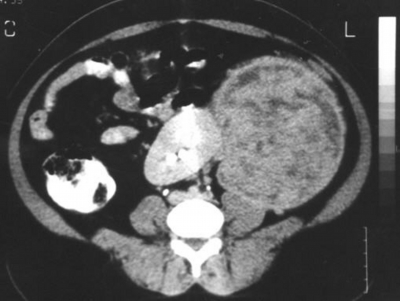
A 44-year-old female patient presented to us with pain and mass in the left lumbar region since 2 months. She also complained of intermittent painless hematuria. She had no other urinary or bowel complaints. She had 2 children and her menstrual history was normal. On examination, a large firm mass with smooth surface was palpable in the left lumbar region that showed hardly any movement with respiration with fullness in renal angle. Urinalysis revealed microscopic hematuria. Serum creatinine was normal. Ultrasonography of the abdomen revealed echogenic mass in relation to the left kidney. Computerised tomography (CT scan) revealed a homogenous mass of 20x 16 cms arising from the lateral aspect of the left kidney. The perirenal tissues and inferior vena cava along with the renal vein appeared to be free from the tumor. (Figure 1) A preoperative diagnosis of exophytic renal cell carcinoma was made. The patient was explored with left paramedian incision with a plan of left radical nephrectomy. The Gerota's fascia was found to be stretched over a huge mass seen arising from the entire aspect of the left kidney. (Figure 2) Left radical nephrectomy was performed. The cut surface of the surgical specimen revealed white whorl like appearance and weighed 3.2 kilograms. Histopathology revealed interlacing bundles of smooth muscles with no mitotic figures and pleomorphism. The tumor was well encapsulated however the origin of the tumor was assumed most probably from the capsule of the kidney. Diagnosis of leiomyoma of the capsule of the left kidney was made. (Figure 3). The patient is totally asymptomatic at 1 year follow-up.
Figure 1
Figure 2
Discussion
Leiomyomatous tumors of the kidney are rare and usually about 4.2-5.2% are discovered on autopsy.2 These slow growing tumors are usually asymptomatic however presentations as large palpable mass (57%), pain (53%) and microscopic hematuria (20%) have also been reported.1 Only one case with gross hematuria has been reported with leiomyoma of the renal pelvis.3 The classical triad for clear cell carcinoma of abdominal pain, mass and hematuria has been encountered in about 3.3% of cases. Constitutional symptoms such as fever, weight loss are not encountered. These tumors are mostly seen between 2nd and 5th decades with median age of 42 and more often in women (66%) and in white race.1 Our patient presented with abdominal mass and gross hematuria and is the second case in literature.
Both the kidneys are equally affected with affinity to the lower pole (74%) of cases. These can be subcapsular, capsular and in the pelvis. Location of these tumors represents the origin of these tumors.1 These tumors usually develop from smooth muscle from the capsule (37%), renal pelvis (17%), renal cortical structure (10%), and indeterminate areas (37%).4 Leiomyomas arising from the smooth muscle from the tunica media of the vessels have not been reported. Leiomyosarcomas are rare malignant counterparts which also originates in same manner of leiomyoma, however it is difficult to distinguish between them clinically.5 There is no relation between the size and the weight of tumor and malignancy.1 Niceta et al have reported leiomyosarcomas manifest with more clinical signs as abdominal mass with pain (67.5%) and gross hematuria (42.5%) along with weight loss and fever.5
On gross pathology, leiomyoma are well encapsulated and well circumscribed while leiomyosarcoma are poorly encapsulated.1 Both have similar whorled appearance with cystic and solid component. Cystic change appears following cystic degeneration and has no correlation with sarcomatous change.1,4 About 17% of renal leiomyomas show hemorrhage and irregular calcification is seen in 20%,6,7 however these do not necessarily suggest benign nature as leiomyosarcomas demonstrate calcification in 10% of cases.5
Microscopic evaluation is the only confirmative method the differentiate between leiomyoma and leiomyosarcoma.7 Presence of mitotic figures, pleomorphism and hyperchromatism reveal malignant change in the interlacing bundles of smooth muscles of leiomyoma of the kidney. Leiomyosarcomas are known to invade the renal vein, inferior vena cava, perirenal fat and also reported to metastasize to peritoneum, omentum, liver, lungs, skin, adrenals, and opposite kidney.1
Ultrasonography, CT scan, arteriography are useful in diagnosis of these tumors.1 Angiographically these tumors may be hypovascular or hypervascular.8 CT scan may at times demonstrate a plane between the kidney and the tumor to suggest it as a capsular leiomyoma. CT scan may demonstrate well circumscribed mass without any extrarenal invasion and its location.1 However only histopathology can differentiate it from exophytic renal cell carcinoma.
Treatment recommended is renal exploration.1 Unroofing is suggested in cystic lesions and partial nephrectomy is done in solid tumors and large cysts. In large tumors, with invasion demonstrated on CT scan radical nephrectomy is advised.1 However, renal leiomyomas are benign and have found to have good postoperative prognosis.
Acknowledgement
We are thankful to the Dean, Dr Kshirsagar for allowing us to publish the hospital data.
Correspondence to
Dr Rege Sameer Ashok C-201, Gagangiri Park Co-op. Hsg. Society, Samata Nagar, Thane. (West) 400606. India. Telephone: 91-22-25385353 Fax: 91-22-24143435 E-mail: samrege@yahoo.com


