Isolated Pancreatic Tuberculosis: A Diagnostic Dilemma With Good Prognosis
G Chiranjiv, O Ashish, V Brinder, T Anuraag
Keywords
pancreas, tuberculosis
Citation
G Chiranjiv, O Ashish, V Brinder, T Anuraag. Isolated Pancreatic Tuberculosis: A Diagnostic Dilemma With Good Prognosis. The Internet Journal of Surgery. 2005 Volume 8 Number 2.
Abstract
Isolated pancreatic tuberculosis is a rare entity even in the endemic areas presenting with a masquerading symptoms of ranging from acute or chronic pancreatitis to carcinoma of pancreas with biliary obstruction to being totally asymptomatic. These cases show good response to antitubercular therapy with a good overall prognosis.
Introduction
Although tuberculosis of gastrointestinal tract is common in developing countries but involvement of pancreas is very rare and has been described in only a handful of case reports.1 In 1944 Auerbach reported that pancreas was affected in 4.7% of cases of miliary tuberculosis.2 We hereby report a case of pancreatic tuberculosis.
Case history
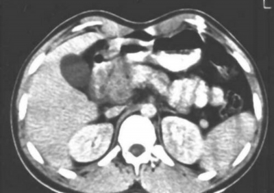
Forty one year male presented with chief complaint of pain in the epigastrium and vomiting for one day. Pain was continuous, noncolicky, moderate intensity, non radiating with no relation to food and posture. Patient had two episodes of bilious vomiting, non projectile in nature .Serum biochemistry showed Total bilirubin to be 6.10 mg/dl, Direct bilirubin as 4.23 mg/dl and Alkaline phosphatase as 1133 IU/L. Serum amylase was 2594 mg/dl and serum lipase was 2537 mg/dl. Contrast enhanced CT scan of abdomen showed a hypodense space occupying lesion (SOL) of 3.3cm in diameter in head of pancreas and few lymph nodes in para-aortic region (Fig 1).
Figure 1
The clinical diagnosis of carcinoma of head pancreas was kept presenting with acute pancreatitis. Fine needle aspiration cytology (F.N.A.C) was done under CT guidance to establish the diagnosis as the presence of para-aortic lymph nodes precluded the curative resection. F.N.A.C. showed it to be tubercular mass (fig 2).
Figure 2
Patient was put on antitubercular drugs (ATT; standard four drug regimen consisting of Isoniazid, Rifampicin, Ethambutol & Pyrazinamide followed at our institute for abdominal tuberculosis) and stenting of the biliary system was deferred. Patient started improving with resolution of jaundice and decrease in the size of mass. Patient was discharged and was on regular follow up. Mass disappeared in 45 days completely on repeat C.E.C.T of abdomen (fig 3). The patient completed the full nine months course of ATT as recommended for abdominal tuberculosis.
Discussion
Clinical involvement of pancreas is rare in case of abdominal tuberculosis as evidenced by a large review of 300 patients with abdominal tuberculosis in India by Bhansali without a single case of clinical involvement of the pancreas.3 So it is a rare entity. The pathogenesis of pancreatic involvement remains obscure; however some authors favor a lympho hematogenous dissemination from a small undetected or reactivated primary or secondary tubercular lesion. The primary focus may be in the intestines or in the retroperitoneal lymph nodes with spread to the pancreas. Head of pancreas is most commonly involved because of its rich lymphatic drainage. Pancreatic tuberculosis generally present with signs of acute or chronic pancreatitis or as biliary obstruction mimicking carcinoma pancreas or a pancreatic abscess.4,5 Patient can present with pain abdomen ,gastrointestinal bleed or may be asymptomatic.6,7 In our case patient presented with complaint of pain abdomen with vomiting and had acute pancreatitis but the probability of pancreatic carcinoma could not be ruled out on radioimaging.
C.E.C.T can show a mass lesion in the pancreas but cannot differentiate between carcinoma and tuberculosis in most of the cases including this case. So F.N.A.C is recommended where tuberculosis is suspected as in the cases of military tuberculosis and immunocompromized patients. We could establish the diagnosis from F.N.A.C. No spillage of tumour cells by F.N.A.C has been reported in carcinoma pancreas so it is a safe procedure and is required to establish the diagnosis of carcinoma pancreas in non operable cases before subjecting the patient to adjuvant therapy.
Correspondence to
Dr Ashish Ohri 3888, street no 2, opp UCO bank, durga puri, haibowal kalan, ludhiana141001 Punjab, India. E-mail – ashohri@yahoo.com


