Colonic Carcinoma in a Young Adult Presenting as an Intussusception
C Quah, V Rajasekaran, R Bryan
Keywords
adult intussusception, atypical abdominal pain, colo-colic intussusception, ct scan, extended right hemicolectomy, hereditary non-polyposis colon cancer, tubulovillous adenoma
Citation
C Quah, V Rajasekaran, R Bryan. Colonic Carcinoma in a Young Adult Presenting as an Intussusception. The Internet Journal of Surgery. 2008 Volume 18 Number 1.
Abstract
Adult intussusception is a rare disease without classical symptoms. A delay in diagnosis may result in a fatal progression to complete bowel obstruction, ischemia, necrosis and perforation. The association of a tumour with intussusception in young adults is rare. We report a case concerning a young lady with a strong family history of bowel cancer presenting with a transverse colon intussusception secondary to colonic carcinoma. A high index of suspicion and an early CT scan was required to prevent delayed diagnosis and the development of complications.
Introduction
Adult intussusception is a rare disease without classical symptoms. It can either be primary or secondary to intestinal pathology with the colo-colic variety being the most common in adults. Although it is a rare entity in the adult, it is important to exclude intussusception as a cause of obstruction or pain, as delay in diagnosis can lead to the development of ischaemic segments of bowel. The association of a tumour with intussusception in young adults is rare. We report a case concerning a young lady with a strong family history of bowel cancer presenting with a transverse colon intussusception secondary to colonic carcinoma. This case highlights the need to consider the diagnosis of intussusception in patients with atypical abdominal pain before ischaemia ensues. For this reason we suggest that a CT scan should be considered early in the management of such patients.
Case Summary
A 32-year-old lady presented with a 15-day history of intermittent abdominal pain and nausea. She had had one episode of diarrhoea with no blood or mucus. Initial examination revealed diffuse tenderness in the epigastrium and left lower quadrant. She subsequently developed tachycardia and increasing abdominal pain which required patient-controlled analgesia. She was found to have normocytic normochromic anaemia. Abdominal plain film revealed a few dilated loops of small bowel. Ultrasound scan was unhelpful. Repeat clinical examination revealed a vague mass in the left upper quadrant of the abdomen. A CT scan was carried out which suggested a large bowel intussusception with a probable mass lesion (figure 1).
Figure 1

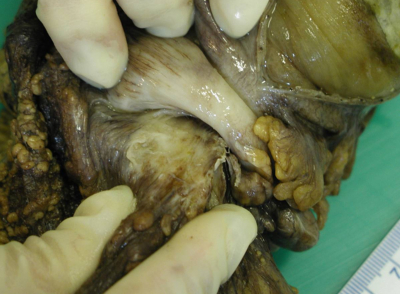
At operation she was found to have a transverse colon intussusception with a tumour just proximal to the splenic flexure (figure 2).
Figure 2
An extended right hemicolectomy was performed. Histology revealed an intussusception around a tubulovillous adenoma with moderately differentiated adenocarcinomatous changes – (Dukes B adenocarcinoma). She was discharged without complications 10 days after surgery.
Following her diagnosis, it emerged that her father was diagnosed with colorectal cancer at 34 years of age and many other family members had been diagnosed with various cancers. She was referred to the regional genetics service who suspects a diagnosis of hereditary non-polyposis colon cancer (HNPCC) and a mutation analysis is currently under way.
Discussion
Intussusception is a rare disease in the adult population characterised by no “classical” abdominal symptoms (1). The diagnosis is elusive and this rare entity needs to be considered in the differential diagnosis of chronic abdominal symptoms in the adult (1). Adult intussusception accounts for about 5% of all intussusceptions (2,5,8) in the Western world, but up to 40% of intussusceptions in tropical Africa (5).
Intussusception occurs when a contracted proximal intestinal segment telescopes into the relaxed distal intestinal segment and normal intestinal peristalsis is disturbed (2). The intussuscepted segment can become markedly swollen and oedematous as a result of circulatory compromise leading to a subsequent risk of necrosis and perforation (2). Colo-colic intussusception is the most common type in adults (6). Common sites of occurrence are the junction between freely mobile segments of bowel and segments that are relatively fixed.
In adults, the majority of cases have a definite underlying cause, with primary intussusceptions accounting for only 15 to 25 percent (1,2,5,7) for most authors. Any intraluminal lesions can act as lead point, which results in a change in the normal peristaltic pattern. Polyps are the most commonly implicated lesions (for example in Peutz-Jeghers syndrome), but several other causes have been described including malignant tumours, Meckel's diverticulum, chronic ulcerations, typhoid enteritis (11), adhesions, worm infestation, endometriosis, mucocele of the appendix, trauma, foreign body, intramural haematomas, previous stoma/anastomotic sites (1), inspissated stools as in cystic fibrosis (9), bezoars (11) and even prolonged fasting (6). Approximately 50% of colonic lesions are due to malignant vegetative neoplasms (3,4,5,8). About 24-25% of the small bowel lesions are malignant (8,11).
Intussusception has no “classical” abdominal symptoms and in adults less than 20 percent of cases present acutely (1). The clinical presentation may include a palpable mass, nausea and vomiting, abdominal colic, change in bowel habit and occult blood per rectum (1). Because of the non-specific nature of these symptoms, the diagnosis is usually delayed. In adults, total obstruction is rare which may account for its chronic symptomatology, while in children, strangulation and gangrene are inevitable features. Spontaneous sloughing of the ischaemic intussuscepted colonic segment has been reported in the literature (5,8,10).
Plain abdominal X-rays are of limited diagnostic value in an adult. Barium enema and ultrasound which are often useful for diagnosis and therapy in children are less helpful in adults. The role of hydrostatic reduction with barium enema remains controversial in adults. Intraluminal seeding or venous embolisation of malignant cells can occur with this procedure (7). CT scan is the primary diagnostic tool of choice as it confirms the diagnosis and may identify the underlying cause (4,5). Surgical resection is the definitive treatment for the invaginating tumour. It is important for the surgeon to take into consideration the clinical status, location and status of the intussusception and condition of the bowel before deciding on whether a primary anastomosis should be performed (3,4). Also manual reduction of the intussusception is not advocated during laparotomy (1,3,8,12).
It is interesting to note that the intussusception caused by the carcinoma in our patient probably resulted in her being diagnosed at a favourable early stage of her disease (Dukes B). Initial investigation by ultrasound was not helpful and delayed her diagnosis. Although no deleterious sequalae resulted from this delay, greater awareness of this condition may have prompted the request for an earlier CT scan leading to a more timely diagnosis and intervention.
Conclusion
In conclusion, adult intussusception is a potentially life-threatening condition which is rare but well recognised. Delay in diagnosis may result in a fatal progression to complete bowel obstruction, ischemia, necrosis and perforation. A high index of suspicion and an early CT scan may prevent delayed diagnosis and the development of complications.
Acknowledgements
1. We would like to thank Dr. Henderson, Consultant Pathologist, for kindly providing the pathology image.
2. Also we would like to thank Mrs. Joan O'Brien, surgical secretary at Friarage Hospital, Northallerton, for her enormous help with the notes of this patient.
Correspondence to
Dr Conal Quah Surgical Trainee Scott Suite Friarage Hospital Northallerton, North Yorkshire DL6 1JG Contact no.: 07738005994 Email: conalquah@yahoo.com