ISPUB.com / IJMB/7/1/11494
  • Author/Editor Login
  • Registration
  • Facebook
  • Google Plus

ISPUB.com

Internet
Scientific
Publications

  • Home
  • Journals
  • Latest Articles
  • Disclaimers
  • Article Submissions
  • Contact
  • Help
  • The Internet Journal of Microbiology
  • Volume 7
  • Number 1

Original Article

UV-B radiation and temperature stress causes variable growth response in Metarhizium anisopliae and Beauveria bassiana isolates

U Mustafa, G Kaur

Keywords

b. bassiana, germination, growth, m. anisopliae, temperature stress, uv-b radiation stress

Citation

U Mustafa, G Kaur. UV-B radiation and temperature stress causes variable growth response in Metarhizium anisopliae and Beauveria bassiana isolates. The Internet Journal of Microbiology. 2008 Volume 7 Number 1.

Abstract

The biocidal effects exerted through the activity of living organisms are practically more feasible and sustainable than the chemical cure. But one of the major threats for the bio-control agents is the on-field exposure to abiotic stresses. In this context, the present in-vitro study was undertaken to investigate the effect of temperature and time-dependent exposure of UV-B radiations on seventeen Beauveria bassiana and fourteen Metarhizium anisopliae isolates. A comparative analysis based on germination, growth and sporulation was undertaken. It was observed that certain isolates showed significant tolerance to these abiotic stresses. In response to UV-B stress, the B. bassiana isolates were more tolerant than the M. anisopliae isolates. But under temperature stress, the M. anisopliae isolates were more tolerant with isolates showing germination, growth and sporulation potential up to 37ºC. Asa conclusion, these stress tolerant entomopathogenic fungal isolates hold high affirmations for commercial market.


Introduction

Commercial biological control involving entomopathogenic fungi in the present global scenario is a hi-tech venture both in terms of safety and sustenance. But the credibility of hi-fidelity management of insect pests by fungus is besotted upon attaining, maintaining and novel sustaining of such fungal strains, in the turmoil multitude of abiotic stresses (1). The continuance of viability and virulence of fungal inoculum (conidia) after field application is the pre-requisite threshold for their efficacy (2). Various isolates of B. bassiana and M. anisopliae have been the most entrusted entomopathogens that have been heavily researched upon and find appreciable on-field commercial usage for insect pest management (3). Upon field application, the entomopathogens are exposed to an array of abiotic stresses like temperature (4), UV radiations (5), humidity-osmolarity (6), edaphic factors and nutrient source (7) that negatively affect the field use of entomopathogens as biocontrol agents. Continued abiotic stress can either have absolute deleterious effect or force natural selection in them (8).

The solar radiation, which includes visible light, ultraviolet radiations, infrared rays and radio waves have been the dominant source in which all organisms evolved and adapted. In biological context, the UV radiations acclaim a special mention in terms of their impact on life (9). Ultraviolet (UV) light is electromagnetic radiation with a wavelength shorter than that of visible light, but longer than soft X-rays. When considering the effect of UV radiations on organisms and the environment, the range of UV wavelength is often subdivided into UV-A or long wave or black light (400-315 nm), UV-B or medium wave (315-280 nm), and UV-C or short wave or germicidal (‹ 280 nm). UV-photons, in particular those belonging to the UV-B type, form covalent bonds between adjacent thymine bases resulting in thymine dimers. Thymine dimers do not base pair normally, which causes distortion of the DNA helix, stalled replication, gaps and misincorporation. These can lead to mutations and ultimately disrupt the normal functioning of the organism (10). Soil temperature is a major factor, which affects the success or failure in the establishment and production of fungal inoculum (11). The entomopathogenic fungi not only have to be tolerant to the soil temperature but also have to survive through thermoregulatory defense response of the host insect (12, 13). It has been demonstrated that stress temperature alters the vegetative growth among isolates of entomopathogenic fungi (14). Dry heat exposure causes DNA damage through base loss leading to depurination and this may cause mutation (15). Wet heat i.e. heat in conjunction with high humidity results in protein denaturation and membrane disorganization. It has been reported that M. anisopliae has temperature tolerance upper limit as 37-40ºC (11). B. bassiana on the other hand can survive up to a maximum temperature of 37 ºC (16). In fungi the temperature range for germination and mycelial growth has been reported to be similar.

The entomopathogenic fungi are natural and cosmopolitan in occurrence. The purpose of this study has been to identify those naturally occurring entomopathogenic fungi, which are naturally resilient to abiotic stresses. With such an objective, in-vitro laboratory studies were undertaken to investigate the stress-tolerant attributes among fourteen M. anisopliae and seventeen B. bassiana isolates. The abiotic stresses, which were studied, were temperature stress and UV-B radiation stress. A comparative analysis based on germination, growth and sporulation parameters was done in case of temperature stress. In UV-B radiation stress studies, only germination assay was undertaken.

Materials and methods
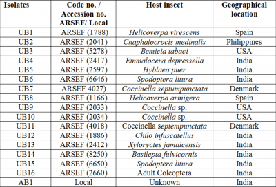
Fungal cultures

The different isolates of the fungus M. anisopliae and B. bassiana were either procured from ARSEF (USDA-ARS Plant Protection Unit) or isolated locally in India. The fourteen isolates of the fungus M. anisopliae were designated as UM1-UM13 and AR1. The seventeen B. bassiana isolates were designated as UB1-UB16 and AB1. The isolates with their accession no., their geographical origin and the host insect from which they were isolated are detailed in Table 1 and Table 2. The isolates were routinely sub cultured on SDA (Sabouraud dextrose Agar) slants at 28°C in incubators and maintained at 4oC.

Figure 1
Table 1. Source of isolates

Figure 2
Table 2. Source of isolates

Colony growth and sporulation (Temperature stress)

Seven day old cultures on SDA slants were used for preparing spore suspension in 0.02% Tween 80 solution at 1x10⁶ spores/ ml. A 200μl of 106 spores were plated on SDA medium and were incubated for 3 days at 28C. At the end of 3 days, 5mm agar disc with mycelia was retrieved with the help of a cork borer and placed in middle of fresh SDA plates (5 replicates/ isolate were maintained) and incubated at 28C, 30C, 34C, 37C and 40C respectively. Radial growth were measured from 3rd day onwards till 8th day Radial growth rate (mmd-1) was calculated from the linear portions of the curves plotted from these values. The plates at 28C served as control. At the end of 8th day, 5mm agar discs were randomly taken with the help of a cork borer. The discs were placed in 10ml of 0.02% (v/v) Tween 80 solution and vortexed to suspend the spores. Spore concentration was determined using a Neubauer Haemocytometer.

Germination (Temperature stress)

Agar slide technique was used for studying the rate of germination. Petri plates were lined with blotting paper discs and 2 glass slides were placed in each of the plates and autoclaved. A 1ml SDA medium was evenly spread on each of the glass slides using micropipette. Conidial suspension was prepared from seven day old cultures with concentrations maintained at 106 conidia/ ml. Approximately 100µl of 106 spores/ml of fungal isolates were spread on the SDA coated slides. The slides were placed back in the Petri plates and the blotting paper discs were moistened with sterilized water. The Petri plates were kept for incubation at 28C, 30C, 34C, 37C and 40C (2 replicates/ isolate/ treatment were maintained). The slides were observed under compound microscope (40X) for germination, every 2 hours, starting from 4th hour after inoculation. A conidium was considered to be germinated when a distinct germ tube projected from it, and was at least twice the diameter of the conidium (17). Approximately 300 conidia were scored per replicate for each of the treatments and the rate of germination determined.

Germination (UV-B radiation stress)

A stock solution of 106 spores/ ml was prepared in a viol and vortexed and divided into 1ml each in 5 polystyrene tubes. The control-experiment tubes were wrapped in aluminum foil (0 hour exposure as the aluminum foil prevented UV-B penetration) and placed together with the test treatment tubes on UV-Trans illuminator, which served as the source of UV-B radiations (320 nm) and gave the desired exposure. Treatments (exposure duration) were 1, 2, 3 and 4 hours exposure to UV-B radiations. Petri plates were lined with blotting paper discs and 2 glass slides were placed in each of the plates and autoclaved. A 1ml SDA medium was evenly spread on each of the glass slides. Approximately 100µl of 106 spores/ml of fungal isolates, which were UV-B treated at different exposure durations, were spread on the SDA coated slides. The slides were placed back in the Petri plates and the blotting paper discs were moistened with sterilized water. The Petri plates were kept for incubation at 28C in dark (2 replicates/ isolate/ treatment were maintained). Observations were taken from 4th hour onwards till 24th hour, and repeated at every 4-hour interval. The slides were observed under compound microscope (40X) for germination. A conidium was considered germinated when a distinct germ tube projected from it. On an average about 300 conidia were scored/ replicate/ treatments and the rate of germination determined.

Statistical analysis

Variances in germination and growth counts among the different treatments and the sample time (days/ hours) were analyzed using procedures for two factor analysis of variance (ANOVA). The data of percentage germination were arc sine percentage square root transformed before analysis.

Results

Colony growth and sporulation (Temperature stress)

The fourteen M. anisopliae isolates showed radial mycelial growth at all the test temperatures except at 40C. All the M. anisopliae isolates showed similar tolerance (P> 0.05) towards temperature stress. But the response of each of the isolates at different temperature showed significant variation (P< 0.05). In M. anisopliae isolates the radial growth was more at 28C but decelerated sharply with increase in temperature at 34C (Table 3). At 37C, the M. anisopliae isolates showed rosette like limited growth which reverted to normal growth pattern on reversion to lower temperature (28C). No growth was observed in plates at 40C and these plates when reverted to lower favorable temperature did not show any resumption of normal growth pattern. All the M. anisopliae isolates showed a 100 fold increase in spore yield at both 28C (control) and 30C but remained practically constant at 34C and 37C, i.e. 106 (the same value as the starting concentration used for spread plating). Isolates UM1, UM2, UM6, UM7 and AR1 showed appreciable growth and sporulation potential at almost all the stress temperatures studied.

The response of different B. bassiana isolates towards temperature stress was significant (P<0.05). All the seven B. bassiana isolates showed mycelial growth at 28C and 30C only and the growth was more at 28C than at 30C (Table 4). Isolates UB2, UB6, UB8, UB9, UB15 and UB16 showed maximum relative radial growth (in comparison to other isolates) at both 28C and 30C. In case of other B. bassiana isolates, the mycelial growth at 30C decreased to an extent with increase in temperature. The B. bassiana isolates showed a 100-fold increase (over the initial concentration of 106 spores/ isolate with which the experiment was started) in spore yield at both 28C (control) and 30C (Table 4). Like mycelial growth, the sporulation in B. bassiana isolates was more at 28C than at 30C. Isolate UB1, UB6 and UB16 showed maximum sporulation (in comparison to other isolates) at 28C and at 30C isolates UB2, UB6, UB8, UB9, UB 15 and UB16 showed highest sporulation compared to other isolates.

Figure 3
Table 3: Temperature dependent growth and sporulation of isolates

Values followed by the same lower case alphabets in the same column are statistically equivalent (P<0.05) according to the Newman-Keul’s multiple range test.

Figure 4
Table 4: Temperature dependent growth and sporulation of isolates

Values followed by the same lower case alphabets in the same column are statistically equivalent (P<0.05) according to the Newman-Keul’s multiple range test.

Germination (Temperature stress)

The effect of temperature on the M. anisopliae isolates was significant (P< 0.05), meaning, that the different isolates exhibited different germination potentiality with varying temperature. Isolates UM2, UM3 and AR1 were the best germinating isolates at all the temperatures studied and had more than 50% of their conidia germinated even at 37C (Table 5). It was observed that the germination rate declined with increase in temperature. Isolates UM6, UM9, UM10, UM12 and UM13 were poor isolates as they did not show any initiation of germination at 8th hour of incubation. None of the isolates could germinate at 40C and when transferred to lower temperatures could not resume normal growth.

The B. bassiana isolates showed good germination at 28C and 30C (Table 6), the germination rate being faster at the former temperature. Isolates UB1 to UB6 and AB1 showed 100% germination at 28C and 30C at 16th hour of incubation. Isolates AB1, UB3 and UB5 showed similar germination response at both 28C and 30C. At 12th hour of incubation at 34C, good pegs (size of pegs and their corresponding spores being almost equal) were seen in almost all the spores of UB5, UB6 and AB1. At 16th hour of incubation at 34C, very good pegs (size of pegs almost twice the size of corresponding spore) were seen in almost all the spores scored, of UB5 and AB1, but these did not germinate further, probably due to the desiccation of the nutrient source on which they were spread. None of the isolates could germinate at 37C and 40C. It was seen that the B. bassiana isolates were equally tolerant (P> 0.05) to temperature stress in context of the germination rate.

Figure 5
Table 5: Temperature dependent percentage germination of isolates

Values followed by the same lower case alphabets in the same column are statistically equivalent (P<0.05) according to the Newman-Keul’s multiple range test.

Figure 6
Table 6: Temperature dependent percentage germination of isolates

Values followed by the same lower case alphabets in the same column are statistically equivalent (P<0.05) according to the Newman-Keul’s multiple range test.

Germination (UV-B radiation stress)

The UV-B dependent germination in M. anisopliae (Table 7) isolates was significant (P< 0.05). The germination potential of each of the isolates showed significant variation (P< 0.05) in context to the UV-B exposure duration i.e. different germination rate at 0 (control), 1, 2, 3 and 4 hours of UV-B exposure. Isolates UM2, UM3 and AR1 were the most robust candidates in response to UV-B radiation stress. Isolates UM6, UM9, UM10, UM12 and UM13 were poor isolates as they did not show any initiation of germination at 8th hour of incubation.

The B. bassiana isolates showed varying tolerance towards UV-B radiation stress and their response was significantly (P<0.05) affected by the dose of UV-B exposure duration. The B. bassiana isolates exhibited appreciable germination at all the four UV-B exposure durations (Table 7) and did not vary much from the control setup (the aluminum foil wrapped tubes). Isolates UB1 to UB7, UB16 and AB1 showed more than 80% effective germination at 1 hour of UV-B exposure at 16th hour of incubation. Isolate AB1 was the most robust candidate in response to UV-B radiation stress and was almost unaffected to changes in UV-B radiation dose. It was generally observed that the germination potential decreased with increase in UV-B radiation dose. Isolate UB5 was almost unaffected at one and two hours of UV-B exposure but showed decline in germination potential at higher exposure durations. Isolate UB7 and UB12-UB15 did not initiate any germination at 8th hour of incubation and so these were considered as poor isolates. Values followed by the same lower case alphabets in the same column are statistically equivalent (P<0.05) according to the Newman-Keul’s multiple range test.

Figure 7
Table 7: UV-B dependent percentage germination of isolates

Values followed by the same lower case alphabets in the same column are statistically equivalent (P<0.05) according to the Newman-Keul’s multiple range test.

Figure 8
Table 8: UV-B dependent percentage germination of isolates

Values followed by the same lower case alphabets in the same column are statistically equivalent (P<0.05) according to the Newman-Keul’s multiple range test.

Discussion

Abiotic stresses, notably extremes in temperature, photon irradiance, water stress and variable concentration of organic-inorganic solutes, frequently limit growth and pathogenic potential of entomopathogenic fungi (18). In addition, more than one abiotic stress can occur at one time. Furthermore, one abiotic stress can decrease an organism’s ability to resist a second stress or vice-versa (8). Great variations in germination, growth and sporulation parameters was observed as isolate response to UV-B radiation and temperature stress, as anticipated from previous reports (19, 4) Certain isolates were appreciably robust in response to these abiotic stresses. Probably the physiology of tolerant isolates is more suited towards abiotic stresses and is thus indicative of their complex genetic base. The tolerant isolates exhibited variation in the phenotypic states depending on the stress stimuli and this capacity is supposed to be adaptive. Phenotypic plasticity exhibited by the entomopathogens was either a reversible or an irreversible plastic response, depending upon the reversion or persistence of the stress stimuli (20, 21). In the present study, it was observed that all M. anisopliae isolates, showed a constant rosette like limited radial growth of 4.5mm at 37C on all the days of observations, and this could revert to normal growth pattern on reversion to lower temperature. On the other hand, B. bassiana isolates which could not show any biological activity at 34C and above exemplifies an irreversible change at either persistence and/ or reversion of temperature stress stimulus.

Abiotic stress tolerance is a polygenically determined attribute. Resilience to abiotic stress by inherent polygenic mechanisms is disadvantageous in terms of a rather complex genetic manipulation strategy for strain improvement. Consequently in vitro assays of abiotic stress inducers, for strain selection (in lieu of strain improvement) offers immediate solutions for identifying robust isolates and considering them for further commercial usage (22). The generalization, and its converse, that an increment in abiotic stress dose (above the condition optima) decrements the biological activities (viz. germination, growth and sporulation) holds true in in- vitro assays on M. anisopliae and B. bassiana isolates. Hedgecock et al., 1995 (23) have reported that temperature is a key factor for conidial viability and longevity of M. anisopliae. James et al., 1998 (24) reported that B. bassiana could grow most rapidly at a continuous temperature of 25-32C, but at temperatures higher than 32±1C, germination was delayed and/ or decreased in some isolates. Results of the present study have shown that the M. anisopliae isolates hold an edge over the B. bassiana isolates for temperature tolerance. The B. bassiana isolates germinated and grew at 28C and 30C; and this is almost consistent with James et al., 1998 (24). All the M. anisopliae isolates exhibited conidial germination, mycelial growth and sporulation up to a maximum temperature of 37C which was 3C more than the tolerance limit of B. bassiana isolates; which is in consistence with the observations of Thomas and Jenkins, 1997 (11).

Studies on UV-B tolerance suggest that with increasing exposure time, the rate of conidial germination declines (5). Fargues et al., 1996 (25) observed that conidia of M. flavoviride were generally more resistant to artificial UV-B irradiance, followed by conidia of B. bassiana, M. anisopliae and Paecilomyces fumosoroseous. In our study, with increase in UV-B exposure duration a corresponding decrease in conidial germination was observed in case of B. bassiana isolates but this decrease was not significant. On the other hand most of the M. anisopliae isolates were susceptible to the increasing dose of UV-B irradiance. It is essential that the entomopathogenic fungi must on priority survive on-field application and also retain activity in the environment. The major impending challenges that the entomopathogens face is survival against fluctuating physical environment. Consequently the artificial in vitro based assays for screening and selection, should adopt an ecological approach that takes into account the features of the environment in which it is intended to be used (26). The northeastern part of India (where this project is currently running) encounters great fluctuations in day-night temperatures and solar radiations. The summer months witness diurnal temperature range of 35-40C, which drops by an amplitude of 5C at night. While during the winter months, the atmospheric temperature varies between 20-25C in the daytime and 10-15C at night. Also during daytime, the sunlight has UV-B component acting in conjunction with the solar temperature. From this study, it can be concluded that B. bassiana isolates are flexible in the sense of application time, as they were not significantly affected by change in UV-B dose. On the other hand, the M. anisopliae isolates could tolerate temperatures from 28C up to 37C and this supports their candidature for application during winter and summer seasons. But as far as their application time is concerned, most isolates can be applied after sunset, thus allowing suitable time for their acclimatization. On coming in contact with the insect cuticle, the most desirable trait for an entomopathogen is its rate of germination which facilitates its growing into the insect haemocoel. As a conclusion of this study, isolate AR1 (M. anisopliae) which showed around 88% germination rate for up to 37C and could tolerate up to 4 hours of UV-B radiation exposure, seems to be a promising candidate for field use.

Acknowledgements

We acknowledge the financial support provided by Department of Science and technology (Project No. SR/ FT/ L-44). We also thank Dr. Richard A. Humber (USDA-ARS) for providing the fungal cultures.

References

1. Tang, Hou. (2001). Effects of environmental factors on virulence of the entomopathogenic fungus, Nomuraea rileyi, against the corn earworm, Helicoverpa armigera (Lep., Noctuidae). J. Appl. Entomol. 125(5), 243–248.
2. Doust, R.A. and Roberts D.W. (1983). Studies on the prolonged storage of Metarhizium anisopliae conidia: Effect of temperature and relative humidity on conidial viability and virulence against mosquitoes. J. Invertbr. Pathol. 41(2), 143-150.
3. Easwaramoorthy, S. (2003). Entomopathogenic Fungi. In: Srivastava RP, ed. Biopesticides and Bioagents in Integrated Pest Management of Agricultural Crops, International Book Distributing Co., India. Pp. 341-379.
4. Rangel, D.E.N. and Braga, G.U.L. Anderson, A.J. and Roberts, D.W. (2005a). Variability in conidial thermo tolerance of Metarhizium anisopliae isolates from different geographic origins. J. Invertbr. Pathol. 88, 116-125.
5. Rangel, D.E.N. Butler, M.J. Torabinejad, J. Anderson, A.J. Braga, G.U.L. Day A.W. and Roberts, D.W. (2006a). Mutants and isolates of Metarhizium anisopliae are diverse in their relationships between conidial pigmentation and stress tolerance. J. Invertbr. Pathol. 93(3), 170-182.
6. Lazzarini, G.M.J. Rocha, L.F.N. and Luz, C. (2006). Impact of moisture on in vitro germination of Metarhizium anisopliae and Beauveria bassiana and their activity on Triatoma infestans. Mycol. Res. 100,485-492.
7. Shah, F.A. Wang, C.S. and Butt, T.M. (2005). Nutrition influences growth and virulence of the insect pathogenic fungus Metarhizium anisopliae. FEMS Microbiol. Letters. 251, 259-266.
8. Rangel, D.E.N. Anderson, A.J. and Roberts, D.W. (2006b). Growth of Metarhizium anisopliae on non-preferred carbon sources yields conidia with increased UV-B tolerance. J. Invertbr. Pathol.93, 127-134.
9. Bjorn, L.O. (2006). Stratospheric ozone, ultraviolet radiation, and cryptogams. Biological Conservation. 135 (3), 326-333.
10. Diffy, B.L. (1991). Solar ultraviolet radiation effects on biological systems. Review in Physics in Medicine and Biology. 36(3), 299-328.
11. Thomas, M.B. and Jenkins, N.E. (1997). Effect of temperature on growth of Metarhizium flavoviride and virulence to the variegated grasshopper, Zonocerus variegatus. Mycol. Res.101 (12), 1469-1474.
12. Inglis, G.D. Jhonson, D.L. and Goettel, M.S. (1996), Effects of Temperature and Thermoregulation on Mycosis by Beauveria bassiana in Grasshoppers. Biol. Control. 7(2), 131-139.
13. Ouedraogo, R.M. Cusson, M. Goettel, M.S. and Brodeur, J. (2003). Inhibition of fungal growth in thermoregulating locusts, Locusta migratoria, infected by the fungus Metarhizium anisopliae var. acridum. J. Invertebr. Pathol. 82,103–109.
14. Ouedraogo, A. Fargues, J. Goettel, M.S. and Lomer, C.J. (2004). Effect of temperature on vegetative growth among isolates of Metarhizium anisopliae and Metarhizium flavoviride. Mycopathologia. 137(1), 37-43.
15. Nicholson, W.L. Munakata, N. Horneck, G. Melosh, H.J. and Setlow, P. (2000). Resistance of Bacillus endospores to extreme terrestrial and extraterrestrial environments. Microbiol. Mol. Biol. Rev. 64, 548–572.
16. Fargues, J. Goettel, M/S. Smits, N. Ouedraogo, A. and Rougier, M. (1997). Effect of temperature on vegetative growth of Beauveria bassiana isolates from different origins. Mycologia, 89, 389–392.
17. Milner, R.J. Huppatz, R.J. and Swaris, S.C. (1991). A new method for assessment of germination of Metarhizium anisopliae conidia. J. Invertbr. Pathol.57, 121-123.
18. Inglis, G.D. Goettel, M.S. Butt, T.M. and Strasser, H. (2001). Use of Hyphomycetous Fungi for Managing Insect Pests. In: Butt TM, Jackson CW, Magan N, Eds. Fungi as Biocontrol Agents, CAB Publishing. Pp.23-69.
19. Braga, G.U.L. Flint, S.D. Miller, C.D. Anderson, A.J. and Roberts, D.W. (2001). Both solar UVA and UVB radiation impair conidial culturability and delay germination in the entomopathogenic fungus Metarhizium anisopliae. Photochem. Photobiol. 74, 734-739.
20. Kassen, R. and Rainey, P.B. (2004). The ecology and genetics of microbial diversity. Annu. Rev. Microbiol. 58,207-231.
21. Rangel, D.E.N. Braga, G.U.L. Anderson, A.J. and Roberts, D.W. (2005b). Influence of growth environment on tolerance to UV-B radiation, germination speed and morphology of Metarhizium anisopliae var. acridium conidia. J. Invertbr. Pathol. 90, 55-58.
22. St. Leger, R. and Screen, S. (2001). Prospects for Strain Improvement of Fungal Pathogens Of Insects and Weeds. In: Butt TM, Jackson CW, Magan N, eds. Fungi as Biocontrol Agents, CAB Publishing, Oxon, pp. 219-237.
23. Hedgecock, S. Moore, D. Higgins, P.M. and Prior, C. (1995). Influence of moisture content on temperature tolerance and storage on Metarhizium flavoviride conidia in an oil formulation. BioControl. Sci. Technol. 5, 371-377.
24. James, R.R. Croft, B.A. Shaffer, B.T. and Lighthart, B. (1998). Impact of temperature and humidity on host-pathogen interactions between Beauveria bassiana and a cocciellid. Environ. Entomol.27, 1506-1513.
25. Fargues, J. Goettel, M.S. Smits, N. Ouedraogo, A. Vidal, C. Lacey, L.L. Lomer, C.J. and Rougier, M. (1996). Variability in susceptibility to simulated sunlight of conidia among isolates of entomopathogenic Hyphomycetes. Mycopathologia. 135,171–181.
26. Deacon, J.W. (1991). Significance of ecology in the development of bio control agents against soil-borne plant pathogens. BioControl Sci. Technol. 1, 5-20.

Author Information

Uzma Mustafa, M.Sc.
Department of Biotechnology, Indian Institute of Technology Guwahati, Guwahati – 39, Assam, India

Gurvinder Kaur, Ph.D.
Department of Biotechnology, Indian Institute of Technology Guwahati, Guwahati – 39, Assam, India

Download PDF

Your free access to ISPUB is funded by the following advertisements:


BACK TO TOP
  • Facebook
  • Google Plus

© 2013 Internet Scientific Publications, LLC. All rights reserved.    UBM Medica Network Privacy Policy