ISPUB.com / IJMB/6/1/9486
  • Author/Editor Login
  • Registration
  • Facebook
  • Google Plus

ISPUB.com

Internet
Scientific
Publications

  • Home
  • Journals
  • Latest Articles
  • Disclaimers
  • Article Submissions
  • Contact
  • Help
  • The Internet Journal of Microbiology
  • Volume 6
  • Number 1

Original Article

Antimicrobial Activity of Eucalyptus major and Eucalyptus baileyana Methanolic Extracts

I Cock

Keywords

antibacterial activity, australian plants, medicinal plants, methanol extracts

Citation

I Cock. Antimicrobial Activity of Eucalyptus major and Eucalyptus baileyana Methanolic Extracts. The Internet Journal of Microbiology. 2008 Volume 6 Number 1.

Abstract

The antimicrobial activity of methanolic extracts of Eucalyptus baileyana leaves and Eucalyptus major leaves and flowers were investigated by disc diffusion assay against a panel of bacteria and fungi. E. baileyana leaf extract inhibited the growth of 6 of the 14 bacteria tested (43%). Gram-positive and Gram-negative bacteria were both inhibited by E. baileyana leaf extract although Gram-positive bacteria were more susceptible. 4 of 11 Gram-negative (36%) and 2 of 3 Gram-positive bacteria (67%) had their growth inhibited by E. baileyana extract. E. major leaf extract displayed broad antibacterial activity, inhibiting the growth of 10 of the 14 bacteria (71%). E. major flower extract inhibited the growth of 7 of the 14 bacteria (50%). E. major leaf and flower extracts both preferentially inhibited Gram-positive bacteria (100%) compared to Gram-negative bacteria (64% inhibition by leaf extract; 36% inhibition by flower extracts). None of the Eucalyptus extracts showed any antifungal activity. The antibacterial activity of E. major leaf extract was further investigated by growth time course assays which showed significant growth inhibition in cultures of Bacillus
cereus and Pseudomonas fluorescens within 1 h and in Bacillus subtilis and Aeromonas hydrophilia within 2 h.


Introduction

Plants produce a wide variety of compounds which in addition to giving them characteristic pigment, odour and flavour characteristics, may also have antimicrobial properties (Cowan, 1999). For thousands of years, traditional plant derived medicines have been used in most parts of the world and their use in fighting microbial disease is becoming the focus of intense study (Bhavnani and Ballow, 2000; Chariandy et al., 1999). Much of the research into traditional medicinal plant use has focused on Asian (Patwardhan et al., 2005), African (Hostettmann et al., 2000) and South American (Paz et al., 1995) plants. Relatively few studies have focused on the antibacterial activity of Australian native plants.

Eucalyptus is a diverse genus of trees in the family Myrtaceae. Of the more than 700 species that comprise this genus, most are endemic to Australia. A smaller number are also native to New Guinea, Indonesia and the Phillipines. Eucalypts can be found in almost every region of the Australian continent. They have also been widely introduced into drier subtropical and tropical regions in areas as diverse as Africa, the Middle East, India, USA and South America. In many of these areas these trees are considered invasive (Santos, 1997) whilst in other areas they are prized for their commercial applications. Eucalypts are valued for their wood and some are also valuable sources of proteins, tannins, gum, and dyes although their most valuable product is the eucalyptus oil that is readily distilled from their leaves (Sartorelli, 2007; Trivedi and Hotchandani, 2004). Essential oils from some Eucalyptus species (eg Eucalyptus pulverulenta) comprise up to 90% cineol (Brophy et al., 1985; Foley and Lassak, 2004). The structure of cineol is shown in figure 1. Essential oils from other plants containing cineol (eg. Heteropyxis natalensis Harv) have been previously demonstrated antimicrobial properties (Gundidza et al., 1993). Eucalyptus oil is used extensively in cleaning and deodourising products as well as in cough drops and decongestants (Sartorelli, 2007). Eucalyptus oil has insect pest repellent properties and is a component in many commercial pesticides (Fradin and Day, 2002).

Figure 1
Figure 1: Chemical structure of cineol (1,3,3-trimethyl- 2- oxabicyclo[2,2,2] octane) the major oil components of leaves.

Australian Eucalyptus species also had a role as traditional bush medicines for Australian Aborigines. Several species have been reported to be used to prepare antisceptic washes (Harborne and Baxter, 1995; Lassak and McCarthy, 2006). The resinous exudate from the trunk of Eucalyptus maculata was also taken internally to cure bladder infections (Lassak and McCarthy, 2006). Oils from several Eucalyptus species have been used for the treatment of upper respiratory tract infections, colds, influenza sinus congestion (Harborne and Baxter, 1995) and pulmonary infections (Low et al, 1974). Many recent studies have reported on the antimicrobial activity of oils from many Eucalyptus species (Sartorelli, 2007; Delaquis et al., 2002; Oyedeji et al., 1999).

The use of essential oils for the testing of antimicrobial activity is not without problems. The relative insolubility of many of the oil components retard their diffusion through agar gels in agar dilution or disc diffusion studies. Many studies have utilised solubilising agents (eg. Tween 80) to aid oil component diffusion, resulting in variable results (Griffin et al., 2000; Hammer et al., 1999). Solubilising agents appear to increase the susceptibility of some bacteria to antimicrobial agents, decrease the susceptibility of others, whilst having no effect on yet other bacteria. A recent study (Cock, 2008) has demonstrated the antibacterial activity of methanolic extracts of Eucalyptus baileyana leaves and Eucalyptus major leaves and flowers against a limited panel of bacteria. The current study was undertaken to validate and extend these observations against a wider panel of bacteria and fungi and to determine minimum inhibitory concentrations (MIC) of these extracts.

Materials and Methods

Plant Collection and Extraction

The extracts investigated in this study have been described previously (Cock, 2008). Briefly, Eucalyptus baileyana (Black Stringybark) leaves and Eucalyptus major (Queensland Grey Gum) leaves and flowers were collected from Toohey Forest, Brisbane, Australia and were identified with reference to a taxonomic key to Toohey Forest plants (Coutts and Catterall, 1980). Samples were dried in a Sunbeam food dehydrator and the dried material was ground to a coarse powder. 1 g of each of the powdered samples was extracted extensively in 50 ml methanol (Ajax, AR grade) for 24 hours at 4 oC with gentle shaking. The extracts were filtered through filter paper (Whatman No. 54) under vacuum followed by drying by rotary evaporation in an Eppendorf concentrator 5301. The resultant pellet was dissolved in 15 ml 20 % methanol. The extract was passed through 0.22 µm filter (Sarstedt) and stored at 4 oC.

Test Microorganisms

All media was supplied by Oxoid Ltd. All microbial strains were obtained from Tarita Morais, Griffith University. Stock cultures of Aeromonas hydrophilia, Alcaligenes feacalis, Bacillus cereus, Bacillus subtilis, Citrobacter freundii, Enterobacter aerogenes, Escherichia coli, Klebsiella pneumoniae, Pseudomonas aeuroginosa, Pseudomonas fluorescens, Salmonella salford, Serratia marcescens, Staphylococcus aureus and Yersinia enterocolitia were subcultured and maintained in nutrient broth at 4 oC. Aspergillus niger, Candida albicans, and Saccharomyces cerevisiae were maintained in Sabouraud media at 4 oC.

Evaluation of Antimicrobial Activity

Antimicrobial activity of each plant extract and was determined using a modified Kirby-Bauer (Bauer et al., 1966) disc diffusion method. Briefly, 100 µl of the test bacteria/fungi were grown in 10 ml of fresh media until they reached a count of approximately 108 cells/ml for bacteria, or 105 cells/ml for fungi. 100 µl of microbial suspension was spread onto agar plates corresponding to the broth in which they were maintained.

The extracts were tested using 5 mm sterilised filter paper discs. Discs were impregnated with 10 µl of the test sample, allowed to dry and placed onto inoculated plates. The plates were allowed to stand at 4 oC for 2 hours before incubation with the test microbial agents. Plates inoculated with A. feacalis, A. hydrophilia, B. cereus, B. subtilis, C. freundii, K. pneumoniae, P. aeuroginosa, P. fluorescens, S. marcescens, Y. enterocolitia, C. albicans and S. cerevisiae were incubated at 30 oC for 24 hours, then the diameters of the inhibition zones were measured in millimetres. Plates inoculated with E. aerogenes, E. coli, S. Salford and S. aureus were incubated at 37 oC for 24 hours, then the diameters of the inhibition zones were measured. A. niger inoculated plates were incubated at 25 oC for 48 hours then the zones of inhibition were measured. All measurements were to the closest whole millimetre. Each antimicrobial assay was performed in at least triplicate. Mean values are reported in this report. Standard discs of ampicillin (2 µg), chloramphenicol (10 µg) or ciprafloxicin (2.5 µg) were obtained from Oxoid Ltd. and served as positive controls for antimicrobial activity. For fungi, nystatin discs (100 µg, Oxoid Ltd.) were used as a positive control. Filter discs impregnated with 10 µl of distilled water were used as a negative control.

Minimum Inhibitory Concentration (MIC) Determination

The minimum inhibitory concentration (MIC) of the plant extracts was determined by the disc diffusion method across a range of doses. The plant extracts were diluted in deionised water across a concentration range of 5 mg/ml to 0.1 mg/ml. Discs were impregnated with 10 µl of the test dilutions, allowed to dry and placed onto inoculated plates. The assay was performed as outlined above and the lowest concentration at which no zone of inhibition was observed was recorded as the MIC.

Bacterial Growth Time Course Assay

3 ml of bacterial cultures (B. cereus, B. subtilis, A. hydrophilia, P. fluorescens) in nutrient broth were added to 27 ml nutrient broth containing 0.5 ml E. major leaf extract (diluted 1 in 10 in sterile deionised water). The tubes were incubated at 30 oC with gentle shaking. The optical density was measured at 550 nm after 0, 1, 2, 4 and 6 h incubations. Control tubes were incubated under the same conditions but without the extract. All assays were performed in triplicate.

Results

E. baileyana leaf extract was diluted to a 14 mg/ml concentration and E. major leaves and flowers were diluted to 28 mg/ml and 35 mg/ml respectively. 10 µl of each extract was tested in the disc diffusion assay against 17 microorganisms (table 1). The E. baileyana leaf extract inhibited the growth of 6 of the 14 bacteria tested (43%). The antibacterial activity was strongest against A. faecalis and S. salford (as determined by the diameter of the zone of inhibition and by MIC (table 2)). E. major leaf and flower extracts were both particularly effective at inhibiting bacterial growth. The leaf extract inhibited the growth of 10 of the 14 bacteria tested (71%) whilst the flower extract inhibited the growth of 7 of the 14 bacteria tested (50%).

Figure 2
Table 1: Antibacterial activity of and extracts.

Numbers indicate the mean diameters of inhibition of triplicate experiments ± standard deviation. – indicates no growth inhibition. Chl indicates chloramphenicol (10 µg) was used as the positive control. Amp indicates ampicillin (2 µg) was used as the positive control. Cip indicates ciprafloxicin (2.5 µg) was used as the positive control. Nys indicates nystatin nystatin discs (100 µg) was used as the positive control.

Both Gram-positive and Gram-negative bacteria were inhibited by E. baileyana leaf extract although Gram-positive bacteria were more susceptible. Of the 11 Gram-negative bacteria tested, 4 (36%) were inhibited by E. baileyana leaf extract. The extract also inhibited the growth of 2 of the 3 Gram-positive bacteria tested (67%). E. major leaf and flower extracts also preferentially inhibited the growth of Gram-positive bacteria. Both E. major leaf and flower extracts inhibited 100% of the Gram-positive bacteria tested. In comparison, E. major leaf and flower extracts inhibited 7 of 14 (50%) and 4 of 14 (29%) of the Gram-negative bacteria tested respectively. None of the Eucalyptus extracts tested displayed any antifungal activity towards any of the fungi tested.

The relative level of antibacterial activity was evaluated by determining the MIC values for each extract against the bacteria which were shown to be susceptible by disc diffusion assays. MICs were evaluated in the current studies by disc diffusion across a range of concentrations. This has previously been determined to be a valid method of MIC determination as MIC values determined by disc diffusion correlate well with those determined by broth dilution assays (Gaudreau et al., 2007). The MIC values determined for these extracts are presented in table 2.

Figure 3
Table 2: Minimum inhibitory concentrations (mg/ml) of and extracts against susceptible bacteria.

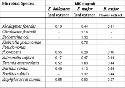
Numbers indicate the mean MIC values of at least least triplicate determinations.

– indicates no growth inhibition.

The antibacterial activity of the E. major leaf extract was further investigated by bacterial growth time course assays in the presence and absence of the extract. The concentration of the extract used in these assays was 46.7 µg/ml. E. major leaf extract was able to significantly inhibit B. cereus (figure 2a), and P. fluorescens (figure 2c) growth within 1 h indicating a rapid antimicrobial action. B. subtilis (figure 2b) and A. hydrophilia (figure 2d) growth was also inhibited by E. major leaf extract, although the inhibition was not significant until 2 h of incubation. Whilst the onset of inhibition of B. subtilis growth was not as rapid as for B. cereus and P. fluorescens, the E. major leaf extract significantly inhibited bacterial growth for the 6 h incubation period. The leaf extract also inhibited growth of A. hydrophilia but this bacteria was able to overcome this inhibitory effect by the end of the 6 h incubation period. After a 6 h incubation the extract treated A. hydrophilia had achieved the same level of growth as the untreated bacteria.

Figure 4
Figure 1: Inhibition of bacterial growth by methanolic extract of leaves against (a) , (b) , (c) , (d) . For all graphs, ? represent measured bacterial growth values for test cultures (with extract) and ? represent control bacterial growth values (no extract). Values are the mean of triplicate determinations.

Discussion

The current study reports on the broad spectrum antimicrobial activity of two Eucalyptus species (E. baileyana and E. major). The ability of these Eucalyptus extracts to inhibit the growth of both Gram-positive and Gram-negative bacteria is in agreement with previous reports of the antibacterial activity of other Eucalyptus species (Sartorelli et al., 2007; Babayi, et al., 2004). These studies also reported the susceptibility of both Gram-positive and Gram-negative bacteria towards various Eucalyptus species extracts. The greater susceptibility of Gram-positive bacteria is in agreement with previously reported results for a wide variety of South American (Paz et al., 1995), African (Kudi et al., 1999; Vlietinck et al., 1995) and Australian (Palombo and Semple, 2001) plant extracts. Results within this laboratory (Cock, 2008) have also confirmed the greater susceptibility of Gram-positive bacteria towards other Australian plant extracts. The Gram-negative bacterial cell wall outer membrane is thought to act as a barrier to many substances including antibiotics (Tortora et al., 2001). The uptake of the Eucalyptus extract antibiotic agents by Gram-negative bacteria is presumably affected by the cell wall outer membrane of some bacteria.

Interestingly, E .major leaf extract displayed no antibacterial activity towards A. hydrophilia in the disc diffusion assays (table 1) although inhibition of A. hydrophilia growth was clearly evident in the bacterial growth assays (figure 2d). The extract appears to inhibit/slow bacterial growth without completely killing all bacteria in the culture. A .hydrophilia growth was initially slower than in the controls, but attained the same level of bacterial growth by the end of the 6 h incubation. It is possible that no inhibition of A. hydrophilia growth was evident in the disc diffusion assays because of the longer incubation time (24 h) required for these assays. Therefore, disc diffusion assays alone may not detect some antimicrobial agents with lower efficiacies because of the incubation time required.

In summary, these studies confirm and extend the previously reported antibacterial activities of E. baileyana and E. major methanolic extracts (Cock, 2008). Most previous studies of Eucalyptus antibacterial activity have reported on the antimicrobial activity of oils (Sartorelli, 2007; Delaquis et al., 2002; Oyedeji et al., 1999) with variable results. The current report uses methanolic extracts to overcome the problems associated with the insolubility of oil components in agar gels. Both Gram-positive and Gram-negative bacteria were susceptible to E. baileyana and E. major extracts. The broad range of microbial susceptibilities indicates the potential of these extracts as a surface disinfectant as well as for medicinal purposes and possibly as food additives to inhibit spoilage. However, further studies are needed before these extracts can be applied to these purposes. In particular, toxicity studies are needed to determine the suitability of these extracts for the use as antiseptic agents and as a food additive.

Aknowledgements

Financial support for this work was provided by the School of Biomolecular and Physical Sciences, Griffith University, Australia.

References

r-0. Babayi H, Kolo I, Okogun JI, Ijah UJ, 2004, The antimicrobial activities of methanolic extracts of Eucalyptus camaldulensis and Terminalia catappa against some pathogenic microorganisms, Biokemistri, 16, 2, 106-111.
r-1. Bauer AW, Kirby WM, Sherris C, Turck M, 1966, Antibiotic susceptibility testing by a standardized single disk method, American Journal of Clinical Pathology, 45, 493-496.
r-2. Bhavnani SM, Ballow CH, 2000, New agents for Gram-positive bacteria, Current Opinions in Microbiology, 3, 528-534.
r-3. Brophy JJ, Lassak EV, Toia RF, 1985, The steam volatile leaf oil of Eucalyptus pulverulenta, Planta Medica, 2, 170-171.
r-4. Chiariandy CM, Seaforth CE, Phelps RH, Pollard GV, Khambay BP, 1999, Screening of medicinal plants from Trinidad and Tobago for antimicrobial and insecticidal properties, Journal of Ethnopharmacology, 64, 265-270.
r-5. Cock IE, 2008, Antibacterial Activity of Selected Australian Native Plant Extracts,
r-6. Internet Journal of Microbiology, 4, 2.
r-7. Coutts RH, Catterall CP, 1980, Identifying the plants of Toohey Forest, Ecos Educational Publishers, Nambour, Australia.
r-8. Cowan MM, 1999, Plant products as antibacterial agents, Clinical Microbiology Reviews, 12, 564-582.
r-9. Delaquis PJ, Stanich K, Girard B, Mazza G, 2002, Antimicrobial activity of individual and mixed fractions of dill, cilantro, coriander and eucalyptus essential oils, International Journal of Food Microbiology, 74, 101-109.
r-10. Foley W, Lassak E, 2004, The potential of bioactive constituents of Eucalyptus foliage as non-wood products from plantations, Publication no. 04/154, Rural Industries and Development Corporation, Australia.
r-11. Fradin MS, Day JF, 2002, Comparitive eficiacy of insect repellants against mosquito bites, New England Journal of Medicine, 347, 1, 13-18.
r-12. Gaudreau C, Girouard Y, Ringuette L, Tsimiklis C, 2007, Comparison of disc diffusion and agar dilution methods for erythromycin and ciprofloxacin susceptibility testing of Campylobacter jejuni subsp. Jejuni, Antimicrobial Agents and Chemotherapy, 1524-1526.
r-13. Griffin SG, Markham JL, Leach DN, 2000, An agar dilution method for the determination of the minimum inhibitory concentration of essential oils, Journal of Essential Oil Research 12, 249-255.
r-14. Gundidza M, Deans SG, Kennedy A, Mavin S, Waten-nam PG, Gray A, 1993, The essential oil from Hetropyxis natalensis Harv: Its antimicrobial activities and phytoconstituents, Journal of the Science of Food and Agriculture, 63, 361-364.
r-15. Hammer KA, Carson CF, Riley TV, 1999, Antimicrobial activity of essential oils and other plant extracts, Journal of Applied Microbiology, 86, 985-990.
r-16. Harborne SB, Baxter H, 1995, Phytochemical dictionary. A handbook of bioactive compounds from plants, Taylor and Francis, London, UK.
r-17. Hostettmann K, Marston A, Ndjoko K, Wolfender J, 2000, The potential of African plants as a source of drugs, Current Organic Chemistry, 4, 973-1010.
r-18. Kudi AC, Umoh JU, Eduvie LO, Gefu J, 1999, Screening of some Nigerian medicinal plants for antibacterial activity, Journal of Ethnopharmacology, 67, 225-228.
r-19. Lassak EV, McCarthy T, 2006, Australian Medicinal Plants, Reed publishers, Australia.
r-20. Low D, Rawal BD, Griffin WJ, 1974, Antibacterial action of the essential oils of some Australian Myrtaceae with special reference to the activity of chromatographic fractions of the oil Eucalyptus citriodora, Planta Medica, 26, 184-189.
r-21. Oyedeji AO, Ekundayo O, Olawore ON, Adeniyi BA, Koenig WA, 1999, Antimicrobial activity of the essential oils of five Eucalyptus species growing in Nigeria, Fitoterapia, 70, 526-528.
r-22. Palombo EA, Semple SJ, 2001, Antibacterial activity of traditional Australian medicinal plants, Journal of Ethnopharmacology, 77, 151-157.
r-23. Patwardhan B, Warude D, Pushpangadan P, Bhatt N, 2005, Ayurveda and traditional Chinese medicine: a comparative overview, EBCAM, 2, 465-473.
r-24. Paz EA, Cerdeiras MP, Fernandez J, Ferreira F, Moyna P, Soubes M, Vazquez A, Vero S, Zunino L, 1995, Screening of Uruguayan medicinal plants for antimicrobial activity, Journal of Ethnopharmacology, 45, 67-70.
r-25. Santos RL, 1997, The Eucalyptus of California. Seeds of good or seeds of evil, Ally-Cass Publications, Denair, California.
r-26. Sartorelli P, Marquioreto AD, Amaral-Baroli A, Lima MEL, Moreno PRH, 2007, Chemical composition and antimicrobial activity of the essential oils from two species of Eucalyptus, Phytotherapy Research, 21, 231-233.
r-27. Tortora GJ, Funke BR, Case CL, 2001, Microbiology: An Introduction, Benjamin Cummings, San Francisco.
r-28. Trivedi NA, Hotchandani SC, 2004, A study of the antimicrobial activity of oil of Eucalyptus, Indian Journal of Pharmacology, 36, 2, 93-95.
r-29. Vlietinck AJ, van Hoof L, Totte J, Lasure A, Vanden Berghe D, Rwangabo PC, Mvukiyumwani J, 1995, Screening of a hundred Rwandese medicinal plants for antimicrobial and antiviral properties, Journal of Ethnopharmacology, 46, 31-47.

Author Information

I.E. Cock
Biomolecular and Physical Sciences, Nathan Campus, Griffith University

Download PDF

Your free access to ISPUB is funded by the following advertisements:


BACK TO TOP
  • Facebook
  • Google Plus

© 2013 Internet Scientific Publications, LLC. All rights reserved.    UBM Medica Network Privacy Policy